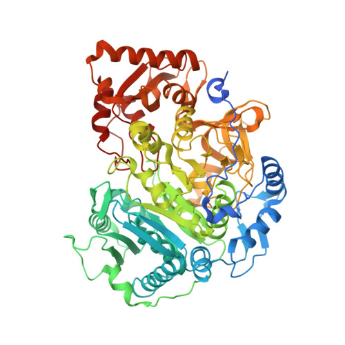
8RWJ

cryoEM structure of Acs1 filament determined by FilamentID


Experimental Data Snapshot

  • Method: ELECTRON MICROSCOPY
  • Resolution: 3.50 Å
  • Aggregation State: FILAMENT 
  • Reconstruction Method: HELICAL 

wwPDB Validation   3D Report Full Report


This is version 1.1 of the entry. See complete history


Literature

FilamentID reveals the composition and function of metabolic enzyme polymers during gametogenesis.

Hugener, J.Xu, J.Wettstein, R.Ioannidi, L.Velikov, D.Wollweber, F.Henggeler, A.Matos, J.Pilhofer, M.

(2024) Cell 187: 3303

  • DOI: https://doi.org/10.1016/j.cell.2024.04.026
  • Primary Citation of Related Structures:  
    8RWJ, 8RWK

  • PubMed Abstract: 

    Gamete formation and subsequent offspring development often involve extended phases of suspended cellular development or even dormancy. How cells adapt to recover and resume growth remains poorly understood. Here, we visualized budding yeast cells undergoing meiosis by cryo-electron tomography (cryoET) and discovered elaborate filamentous assemblies decorating the nucleus, cytoplasm, and mitochondria. To determine filament composition, we developed a "filament identification" (FilamentID) workflow that combines multiscale cryoET/cryo-electron microscopy (cryoEM) analyses of partially lysed cells or organelles. FilamentID identified the mitochondrial filaments as being composed of the conserved aldehyde dehydrogenase Ald4 ALDH2 and the nucleoplasmic/cytoplasmic filaments as consisting of acetyl-coenzyme A (CoA) synthetase Acs1 ACSS2 . Structural characterization further revealed the mechanism underlying polymerization and enabled us to genetically perturb filament formation. Acs1 polymerization facilitates the recovery of chronologically aged spores and, more generally, the cell cycle re-entry of starved cells. FilamentID is broadly applicable to characterize filaments of unknown identity in diverse cellular contexts.


  • Organizational Affiliation

    Institute of Molecular Biology and Biophysics, ETH Zürich, 8093 Zürich, Switzerland; Institute of Biochemistry, ETH Zürich, 8093 Zürich, Switzerland; Max Perutz Labs, University of Vienna, 1030 Vienna, Austria.


Macromolecules
Find similar proteins by:  (by identity cutoff)  |  3D Structure
Entity ID: 1
MoleculeChains Sequence LengthOrganismDetailsImage
Acetyl-coenzyme A synthetase713Saccharomyces cerevisiae SK1Mutation(s): 0 
EC: 6.2.1.1
UniProt
Find proteins for N1P7N2 (Saccharomyces cerevisiae (strain CEN.PK113-7D))
Explore N1P7N2 
Go to UniProtKB:  N1P7N2
Entity Groups  
Sequence Clusters30% Identity50% Identity70% Identity90% Identity95% Identity100% Identity
UniProt GroupN1P7N2
Sequence Annotations
Expand
  • Reference Sequence
Small Molecules
Ligands 1 Unique
IDChains Name / Formula / InChI Key2D Diagram3D Interactions
6R9 (Subject of Investigation/LOI)
Query on 6R9

Download Ideal Coordinates CCD File 
J [auth D]
K [auth I]
L [auth A]
M [auth B]
N [auth C]
J [auth D],
K [auth I],
L [auth A],
M [auth B],
N [auth C],
O [auth E],
P [auth F],
Q [auth G],
R [auth H]
[[(2~{R},3~{S},4~{R},5~{R})-5-(6-aminopurin-9-yl)-3,4-bis(oxidanyl)oxolan-2-yl]methoxy-oxidanyl-phosphoryl] ethanoate
C12 H16 N5 O8 P
UBPVOHPZRZIJHM-WOUKDFQISA-N
Experimental Data & Validation

Experimental Data

  • Method: ELECTRON MICROSCOPY
  • Resolution: 3.50 Å
  • Aggregation State: FILAMENT 
  • Reconstruction Method: HELICAL 
EM Software:
TaskSoftware PackageVersion
MODEL REFINEMENTPHENIX1.14_3260:
RECONSTRUCTIONRELION

Structure Validation

View Full Validation Report


Entry History & Funding Information

Deposition Data


Funding OrganizationLocationGrant Number
Swiss National Science FoundationSwitzerland176108
European Research Council (ERC)European Union101002629

Revision History  (Full details and data files)

  • Version 1.0: 2024-06-26
    Type: Initial release
  • Version 1.1: 2024-07-03
    Changes: Data collection, Database references