Enhanced stabilisation and reduced fibril forming potential of an amyloidogenic light chain using a variable heavy domain to mimic the homodimer complex.
Maerivoet, A., Price, R., Galmiche, C., Scott-Tucker, A., Kennedy, J., Crabbe, T., Antonyuk, S., Madine, J.(2024) FEBS J 291: 4913-4929
- PubMed: 38982771 Search on PubMed
- DOI: https://doi.org/10.1111/febs.17223
- Primary Citation Related Structures:
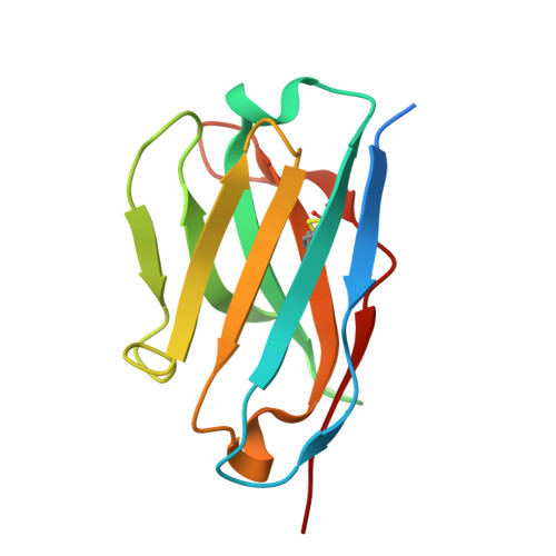
8RPD, 8RPE - PubMed Abstract:
Light chain amyloidosis (AL), is classified as a plasma cell dyscrasia, whereby a mutant plasma cell multiplies uncontrollably and secretes enormous amounts of immunoglobulin-free light chain (FLC) fragments. These FLCs undergo a process of misfolding and aggregation into amyloid fibrils, that can cause irreversible system-wide damage. Current treatments that focus on depleting the underlying plasma cell clone are often poorly tolerated, particularly in patients with severe cardiac involvement, meaning patient prognosis is poor. An alternative treatment approach currently being explored is the inhibition of FLC aggregation by stabilisation of the native conformer. Here, we aimed to identify and characterise antibody fragments that target FLC domains and promote their stabilisation. Using phage-display screening methods, we identified a variable heavy (VH) domain, termed VH1, targeted towards the FLC. Using differential scanning fluorimetry and surface plasmon resonance, VH1 was characterised to bind and kinetically stabilise an amyloidogenic FLC, whereby a > 5.5 °C increase in thermal stability was noted. This improved stability corresponded to the inhibition of fibril formation, where 10 : 1 LC : VH1 concentration reduced aggregation to baseline levels. X-ray crystallographic structures of the LC : VH1 complex at atomic resolution revealed binding in a 1 : 1 ratio, mimicking the dimeric antigen binding sites of the native immunoglobulin molecule and the native LC homodimer.
- Institute of Systems, Molecular and Integrative Biology, University of Liverpool, UK.
Organizational Affiliation: