8RKQ

Structure of human DELTA-1-PYRROLINE-5-CARBOXYLATE DEHYDROGENASE (ALDH4A1) complexed with the molecular tweezer CLR01


Experimental Data Snapshot

  • Method: X-RAY DIFFRACTION
  • Resolution: 2.60 Å
  • R-Value Free: 0.277 
  • R-Value Work: 0.231 
  • R-Value Observed: 0.232 

wwPDB Validation   3D Report Full Report


Ligand Structure Quality Assessment 


This is version 1.0 of the entry. See complete history


Literature

How Do Molecular Tweezers Bind to Proteins? Lessons from X-ray Crystallography.

Porfetye, A.T.Stege, P.Rebollido-Rios, R.Hoffmann, D.Schrader, T.Vetter, I.R.

(2024) Molecules 29

  • DOI: https://doi.org/10.3390/molecules29081764
  • Primary Citation of Related Structures:  
    8RKQ, 8RKR

  • PubMed Abstract: 

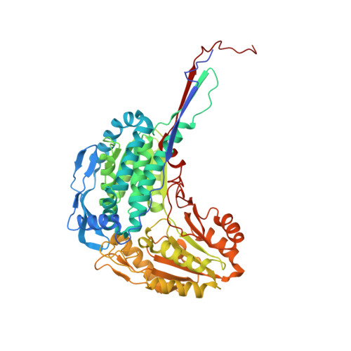
    To understand the biological relevance and mode of action of artificial protein ligands, crystal structures with their protein targets are essential. Here, we describe and investigate all known crystal structures that contain a so-called "molecular tweezer" or one of its derivatives with an attached natural ligand on the respective target protein. The aromatic ring system of these compounds is able to include lysine and arginine side chains, supported by one or two phosphate groups that are attached to the half-moon-shaped molecule. Due to their marked preference for basic amino acids and the fully reversible binding mode, molecular tweezers are able to counteract pathologic protein aggregation and are currently being developed as disease-modifying therapies against neurodegenerative diseases such as Alzheimer's and Parkinson's disease. We analyzed the corresponding crystal structures with 14-3-3 proteins in complex with mono- and diphosphate tweezers. Furthermore, we solved crystal structures of two different tweezer variants in complex with the enzyme Δ 1 -Pyrroline-5-carboxyl-dehydrogenase (P5CDH) and found that the tweezers are bound to a lysine and methionine side chain, respectively. The different binding modes and their implications for affinity and specificity are discussed, as well as the general problems in crystallizing protein complexes with artificial ligands.


  • Organizational Affiliation

    Department of Mechanistic Cell Biology, Max-Planck Institute of Molecular Physiology, Otto-Hahn-Straße 11, 44227 Dortmund, Germany.


Macromolecules
Find similar proteins by:  (by identity cutoff)  |  3D Structure
Entity ID: 1
MoleculeChains Sequence LengthOrganismDetailsImage
Delta-1-pyrroline-5-carboxylate dehydrogenase, mitochondrial
A, B, C, D
567Homo sapiensMutation(s): 0 
Gene Names: ALDH4A1ALDH4P5CDH
EC: 1.2.1.88
UniProt & NIH Common Fund Data Resources
Find proteins for P30038 (Homo sapiens)
Explore P30038 
Go to UniProtKB:  P30038
PHAROS:  P30038
GTEx:  ENSG00000159423 
Entity Groups  
Sequence Clusters30% Identity50% Identity70% Identity90% Identity95% Identity100% Identity
UniProt GroupP30038
Sequence Annotations
Expand
  • Reference Sequence
Small Molecules
Ligands 1 Unique
IDChains Name / Formula / InChI Key2D Diagram3D Interactions
9SZ (Subject of Investigation/LOI)
Query on 9SZ

Download Ideal Coordinates CCD File 
E [auth A](1R,5S,9S,16R,20R,24S,28S,35R)-3,22-Bis(dihydroxyphosphoryloxy)tridecacyclo[22.14.1.15,20.19,16.128,35.02,23.04,21.06,19.08,17.010,15.025,38.027,36.029,34]dotetraconta-2(23),3,6,8(17),10,12,14,18,21,25,27(36),29,31,33,37-pentadecaene
C42 H32 O8 P2
BZESKQAEKFQSPD-FCODNRGNSA-N
Experimental Data & Validation

Experimental Data

  • Method: X-RAY DIFFRACTION
  • Resolution: 2.60 Å
  • R-Value Free: 0.277 
  • R-Value Work: 0.231 
  • R-Value Observed: 0.232 
  • Space Group: P 65
Unit Cell:
Length ( Å )Angle ( ˚ )
a = 148.752α = 90
b = 148.752β = 90
c = 189.957γ = 120
Software Package:
Software NamePurpose
REFMACrefinement
XSCALEdata scaling
PDB_EXTRACTdata extraction
XDSdata reduction
PHASERphasing

Structure Validation

View Full Validation Report


Ligand Structure Quality Assessment 


Entry History & Funding Information

Deposition Data


Funding OrganizationLocationGrant Number
German Research Foundation (DFG)GermanyCRC1093

Revision History  (Full details and data files)

  • Version 1.0: 2024-05-08
    Type: Initial release