8RCO | pdb_00008rco

Structure of Human Serum Albumin in complex with Aristolochic Acid II at 1.9 A resolution


Experimental Data Snapshot

  • Method: X-RAY DIFFRACTION
  • Resolution: 1.90 Å
  • R-Value Free: 
    0.271 (Depositor), 0.271 (DCC) 
  • R-Value Work: 
    0.230 (Depositor), 0.231 (DCC) 
  • R-Value Observed: 
    0.232 (Depositor) 

Starting Model: experimental
View more details

wwPDB Validation 3D Report Full Report

Validation slider image for 8RCO

Ligand Structure Quality Assessment 


This is version 1.2 of the entry. See complete history

Literature

Structural and mechanistic insights into the transport of aristolochic acids and their active metabolites by human serum albumin.

Pomyalov, S.Minetti, C.A.Remeta, D.P.Bonala, R.Johnson, F.Zaitseva, I.Iden, C.Golebiewska, U.Breslauer, K.J.Shoham, G.Sidorenko, V.S.Grollman, A.P.

(2024) J Biological Chem 300: 107358-107358

  • DOI: https://doi.org/10.1016/j.jbc.2024.107358
  • Primary Citation Related Structures: 
    8RCO, 8RCP, 8RGK, 8RGL

  • PubMed Abstract: 

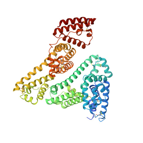
    Aristolochic acids I and II (AA-I/II) are carcinogenic principles of Aristolochia plants, which have been employed in traditional medicinal practices and discovered as food contaminants. While the deleterious effects of AAs are broadly acknowledged, there is a dearth of information to define the mechanisms underlying their carcinogenicity. Following bioactivation in the liver, N-hydroxyaristolactam and N-sulfonyloxyaristolactam metabolites are transported via circulation and elicit carcinogenic effects by reacting with cellular DNA. In this study, we apply DNA adduct analysis, X-ray crystallography, isothermal titration calorimetry (ITC) and fluorescence quenching to investigate the role of human serum albumin (HSA) in modulating AA carcinogenicity. We find that HSA extends the half-life and reactivity of N-sulfonyloxyaristolactam-I with DNA, thereby protecting activated AAs from heterolysis. Applying novel pooled plasma HSA crystallization methods, we report high-resolution structures of myristic acid-enriched HSA (HSA MYR ) and its AA complexes (HSA MYR /AA-I and HSA MYR /AA-II) at 1.9 Å resolution. Whereas AA-I is located within HSA subdomain IB, AA-II occupies subdomains IIA and IB. ITC binding profiles reveal two distinct AA sites in both complexes with association constants of 1.5 and 0.5 · 10 6 M -1 for HSA/AA-I versus 8.4 and 9.0 · 10 5 M -1 for HSA/AA-II. Fluorescence quenching of the HSA Trp 214 suggests variable impacts of fatty acids on ligand binding affinities. Collectively, our structural and thermodynamic characterizations yield significant insights into AA binding, transport, toxicity, and potential allostery, critical determinants for elucidating the mechanistic roles of HSA in modulating AA carcinogenicity.


  • Organizational Affiliation
    • Institute of Chemistry, The Hebrew University of Jerusalem, Edmond J. Safra Campus - Givat Ram, Jerusalem 91904 Israel.

Macromolecule Content 

  • Total Structure Weight: 143.17 kDa 
  • Atom Count: 10,053 
  • Modeled Residue Count: 1,164 
  • Deposited Residue Count: 1,218 
  • Unique protein chains: 1

Macromolecules

Find similar proteins by:|  3D Structure
Entity ID: 1
MoleculeChains  Sequence LengthOrganismDetailsImage
Serum albumin
A, B
609Homo sapiensMutation(s): 0 
UniProt & NIH Common Fund Data Resources
Find proteins for P02768 (Homo sapiens)
Explore P02768 
Go to UniProtKB:  P02768
PHAROS:  P02768
GTEx:  ENSG00000163631 
Entity Groups
Sequence Clusters30% Identity50% Identity70% Identity90% Identity95% Identity100% Identity
UniProt GroupP02768
Sequence Annotations
Expand
Reference Sequence

Small Molecules

Ligands 3 Unique
IDChains Name / Formula / InChI Key2D Diagram3D Interactions
GOR
(Subject of Investigation/LOI)

Query on GOR


Download:Ideal Coordinates CCD File
L [auth A],
M [auth A],
U [auth B],
V [auth B]
6-nitronaphtho[1,2-e][1,3]benzodioxole-5-carboxylic acid
C16 H9 N O6
MEEXETVZNQYRSP-UHFFFAOYSA-N
MYR

Query on MYR


Download:Ideal Coordinates CCD File
C [auth A]
D [auth A]
E [auth A]
F [auth A]
G [auth A]
C [auth A],
D [auth A],
E [auth A],
F [auth A],
G [auth A],
H [auth A],
N [auth B],
O [auth B],
P [auth B],
Q [auth B],
R [auth B],
S [auth B]
MYRISTIC ACID
C14 H28 O2
TUNFSRHWOTWDNC-UHFFFAOYSA-N
EDO

Query on EDO


Download:Ideal Coordinates CCD File
I [auth A],
J [auth A],
K [auth A],
T [auth B]
1,2-ETHANEDIOL
C2 H6 O2
LYCAIKOWRPUZTN-UHFFFAOYSA-N

Experimental Data & Validation

Experimental Data

  • Method: X-RAY DIFFRACTION
  • Resolution: 1.90 Å
  • R-Value Free:  0.271 (Depositor), 0.271 (DCC) 
  • R-Value Work:  0.230 (Depositor), 0.231 (DCC) 
  • R-Value Observed: 0.232 (Depositor) 
Space Group: P 1 21 1
Unit Cell:
Length ( Å )Angle ( ˚ )
a = 94.821α = 90
b = 38.671β = 104.926
c = 180.841γ = 90
Software Package:
Software NamePurpose
PHENIXrefinement
DIALSdata reduction
Aimlessdata scaling
PHENIXphasing

Structure Validation

View Full Validation Report


Ligand Structure Quality Assessment 


Entry History 

& Funding Information

Deposition Data


Funding OrganizationLocationGrant Number
Not fundedIsrael--

Revision History  (Full details and data files)

  • Version 1.0: 2024-06-26
    Type: Initial release
  • Version 1.1: 2024-07-03
    Changes: Database references
  • Version 1.2: 2024-10-23
    Changes: Structure summary