8RBJ | pdb_00008rbj

Structure of fungal tRNA ligase in complex with RNA


Experimental Data Snapshot

  • Method: X-RAY DIFFRACTION
  • Resolution: 2.37 Å
  • R-Value Free: 
    0.218 (Depositor), 0.217 (DCC) 
  • R-Value Work: 
    0.178 (Depositor), 0.179 (DCC) 
  • R-Value Observed: 
    0.180 (Depositor) 

Starting Model: experimental
View more details

wwPDB Validation   3D Report Full Report


Ligand Structure Quality Assessment 


This is version 1.2 of the entry. See complete history


Literature

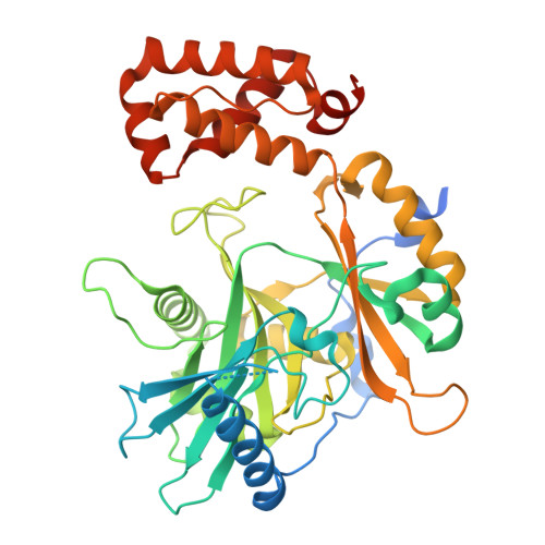
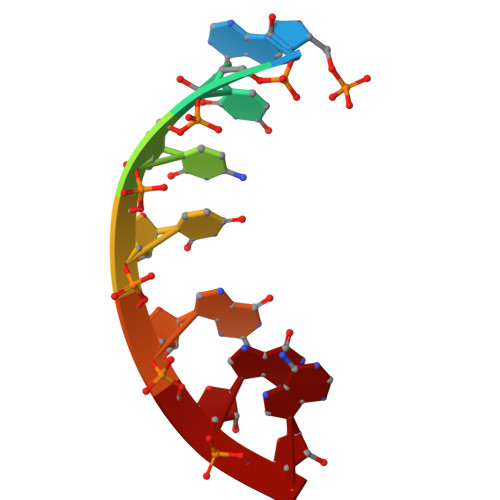
Structure of fungal tRNA ligase Trl1 with RNA reveals conserved substrate-binding principles.

Kohler, S.Kopp, J.Maiti, S.Bujnicki, J.M.Peschek, J.

(2025) Nat Struct Mol Biol 32: 1657-1668

  • DOI: https://doi.org/10.1038/s41594-025-01589-3
  • Primary Citation of Related Structures:  
    8RBJ

  • PubMed Abstract: 

    RNA ligases play a vital role in RNA processing and maturation, including tRNA splicing, RNA repair and the unfolded protein response (UPR). In fungi and plants, the tripartite tRNA ligase Trl1 catalyzes the joining of TSEN-cleaved pre-tRNA exon halves. Trl1 also functions as ligase in the non-conventional HAC1 mRNA splicing during the UPR. The final ligation step is performed by the N-terminal adenylyltransferase domain (ligase; LIG). The spatial arrangement of the exon ends during the ligation reaction has remained elusive. Here we report the crystal structure of Chaetomium thermophilum Trl1-LIG in complex with a tRNA-derived substrate. Our structure represents a snapshot of the activated RNA intermediate and defines the conserved substrate-binding interface. The underlying enzyme-substrate interplay reveals a substrate-binding principle shared by adenylyltransferases. Moreover, we identify the determinants of RNA end specificity as well as the specific roles of Trl1-LIG's subdomains during ligase activation, substrate binding and phosphoryl transfer.


  • Organizational Affiliation
    • Heidelberg University, Biochemistry Center (BZH), Heidelberg, Germany.

Macromolecules

Find similar proteins by:  (by identity cutoff)  |  3D Structure
Entity ID: 1
MoleculeChains Sequence LengthOrganismDetailsImage
tRNA ligase434Thermochaetoides thermophilaMutation(s): 0 
Gene Names: CTHT_0034810
EC: 6.5.1.3
UniProt
Find proteins for G0S6G2 (Chaetomium thermophilum (strain DSM 1495 / CBS 144.50 / IMI 039719))
Explore G0S6G2 
Go to UniProtKB:  G0S6G2
Entity Groups  
Sequence Clusters30% Identity50% Identity70% Identity90% Identity95% Identity100% Identity
UniProt GroupG0S6G2
Sequence Annotations
Expand
  • Reference Sequence

Find similar nucleic acids by:  Sequence   |   3D Structure  

Entity ID: 2
MoleculeChains LengthOrganismImage
RNAB [auth C],
C [auth B]
7Saccharomyces cerevisiae
Sequence Annotations
Expand
  • Reference Sequence
Small Molecules
Ligands 1 Unique
IDChains Name / Formula / InChI Key2D Diagram3D Interactions
AMP (Subject of Investigation/LOI)
Query on AMP

Download Ideal Coordinates CCD File 
D [auth B]ADENOSINE MONOPHOSPHATE
C10 H14 N5 O7 P
UDMBCSSLTHHNCD-KQYNXXCUSA-N
Experimental Data & Validation

Experimental Data

  • Method: X-RAY DIFFRACTION
  • Resolution: 2.37 Å
  • R-Value Free:  0.218 (Depositor), 0.217 (DCC) 
  • R-Value Work:  0.178 (Depositor), 0.179 (DCC) 
  • R-Value Observed: 0.180 (Depositor) 
Space Group: P 21 21 21
Unit Cell:
Length ( Å )Angle ( ˚ )
a = 51.56α = 90
b = 75.36β = 90
c = 124.56γ = 90
Software Package:
Software NamePurpose
XDSdata reduction
Aimlessdata scaling
PHASERphasing
PHENIXrefinement

Structure Validation

View Full Validation Report


Ligand Structure Quality Assessment 


Entry History & Funding Information

Deposition Data


Funding OrganizationLocationGrant Number
German Research Foundation (DFG)Germany442512666
German Research Foundation (DFG)Germany439669440

Revision History  (Full details and data files)

  • Version 1.0: 2025-06-18
    Type: Initial release
  • Version 1.1: 2025-07-30
    Changes: Database references
  • Version 1.2: 2025-10-01
    Changes: Database references