8R8V | pdb_00008r8v

Human PADI4 in complex with cyclic peptide PADI4_11


Experimental Data Snapshot

  • Method: ELECTRON MICROSCOPY
  • Resolution: 3.60 Å
  • Aggregation State: PARTICLE 
  • Reconstruction Method: SINGLE PARTICLE 

wwPDB Validation 3D Report Full Report

Validation slider image for 8R8V

This is version 1.1 of the entry. See complete history

Literature

A cyclic peptide toolkit reveals mechanistic principles of peptidylarginine deiminase IV regulation.

Bertran, M.T.Walmsley, R.Cummings, T.Aramburu, I.V.Benton, D.J.Mora Molina, R.Assalaarachchi, J.Chasampalioti, M.Swanton, T.Joshi, D.Federico, S.Okkenhaug, H.Yu, L.Oxley, D.Walker, S.Papayannopoulos, V.Suga, H.Christophorou, M.A.Walport, L.J.

(2024) Nat Commun 15: 9746-9746

  • DOI: https://doi.org/10.1038/s41467-024-53554-1
  • Primary Citation Related Structures: 
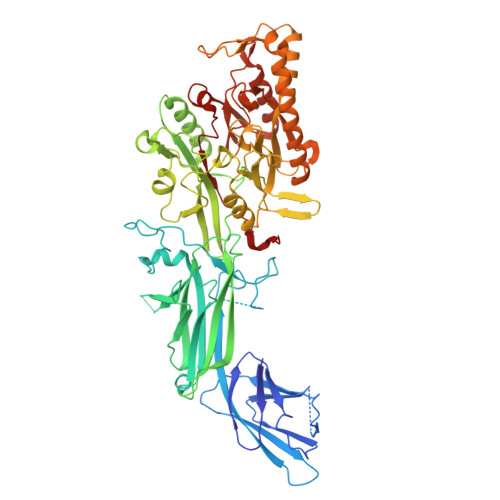
    8R8U, 8R8V

  • PubMed Abstract: 

    Peptidylarginine deiminase IV (PADI4, PAD4) deregulation promotes the development of autoimmunity, cancer, atherosclerosis and age-related tissue fibrosis. PADI4 additionally mediates immune responses and cellular reprogramming, although the full extent of its physiological roles is unexplored. Despite detailed molecular knowledge of PADI4 activation in vitro, we lack understanding of its regulation within cells, largely due to a lack of appropriate systems and tools. Here, we develop and apply a set of potent and selective PADI4 modulators. Using the mRNA-display-based RaPID system, we screen >10 12 cyclic peptides for high-affinity, conformation-selective binders. We report PADI4_3, a cell-active inhibitor specific for the active conformation of PADI4; PADI4_7, an inert binder, which we functionalise for the isolation and study of cellular PADI4; and PADI4_11, a cell-active PADI4 activator. Structural studies with PADI4_11 reveal an allosteric binding mode that may reflect the mechanism that promotes cellular PADI4 activation. This work contributes to our understanding of PADI4 regulation and provides a toolkit for the study and modulation of PADI4 across (patho)physiological contexts.


  • Organizational Affiliation
    • Protein-Protein Interaction Laboratory, The Francis Crick Institute, London, NW1 1AT, UK.

Macromolecule Content 

  • Total Structure Weight: 153.12 kDa 
  • Atom Count: 10,202 
  • Modeled Residue Count: 1,294 
  • Deposited Residue Count: 1,366 
  • Unique protein chains: 2

Macromolecules

Find similar proteins by:|  3D Structure
Entity ID: 1
MoleculeChains  Sequence LengthOrganismDetailsImage
Protein-arginine deiminase type-4A,
D [auth B]
670Homo sapiensMutation(s): 3 
Gene Names: PADI4
EC: 3.5.3.15
UniProt & NIH Common Fund Data Resources
Find proteins for Q9UM07 (Homo sapiens)
Explore Q9UM07 
Go to UniProtKB:  Q9UM07
PHAROS:  Q9UM07
GTEx:  ENSG00000159339 
Entity Groups
Sequence Clusters30% Identity50% Identity70% Identity90% Identity95% Identity100% Identity
UniProt GroupQ9UM07
Sequence Annotations
Expand
Reference Sequence
Find similar proteins by:  Sequence   |   3D Structure  
Entity ID: 2
MoleculeChains  Sequence LengthOrganismDetailsImage
Cyclic Peptide PADI4_11B [auth C],
C [auth D]
13synthetic constructMutation(s): 0 

Small Molecules

Ligands 1 Unique
IDChains Name / Formula / InChI Key2D Diagram3D Interactions
CA

Query on CA


Download:Ideal Coordinates CCD File
E [auth A]
F [auth A]
G [auth A]
H [auth A]
I [auth A]
E [auth A],
F [auth A],
G [auth A],
H [auth A],
I [auth A],
J [auth B],
K [auth B],
L [auth B],
M [auth B],
N [auth B]
CALCIUM ION
Ca
BHPQYMZQTOCNFJ-UHFFFAOYSA-N

Experimental Data & Validation

Experimental Data

  • Method: ELECTRON MICROSCOPY
  • Resolution: 3.60 Å
  • Aggregation State: PARTICLE 
  • Reconstruction Method: SINGLE PARTICLE 
EM Software:
TaskSoftware PackageVersion
MODEL REFINEMENTPHENIX

Structure Validation

View Full Validation Report


Entry History 

& Funding Information

Deposition Data


Funding OrganizationLocationGrant Number
The Francis Crick InstituteUnited KingdomCC2030
The Francis Crick InstituteUnited KingdomFC0010129
The Francis Crick InstituteUnited KingdomFC001134

Revision History  (Full details and data files)

  • Version 1.0: 2024-11-27
    Type: Initial release
  • Version 1.1: 2025-07-09
    Changes: Data collection