8R7U | pdb_00008r7u

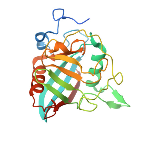
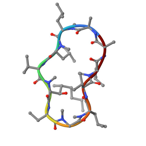
Crystal Structure of Cyclophilin TgCyp23 from Toxoplasma gondii in complex with dihydro Cyclosporin A


Experimental Data Snapshot

  • Method: X-RAY DIFFRACTION
  • Resolution: 1.20 Å
  • R-Value Free: 
    0.187 (Depositor), 0.192 (DCC) 
  • R-Value Work: 
    0.172 (Depositor), 0.181 (DCC) 
  • R-Value Observed: 
    0.172 (Depositor) 

Starting Model: experimental
View more details

wwPDB Validation 3D Report Full Report

Validation slider image for 8R7U

This is version 1.0 of the entry. See complete history. 

Literature

Evaluating the potential of non-immunosuppressive cyclosporin analogs for targeting Toxoplasma gondii cyclophilin: Insights from structural studies.

Favretto, F., Jimenez-Faraco, E., Catucci, G., Di Matteo, A., Travaglini-Allocatelli, C., Sadeghi, S.J., Dominici, P., Hermoso, J.A., Astegno, A.

(2024) Protein Sci 33: e5157-e5157

  • DOI: https://doi.org/10.1002/pro.5157
  • Primary Citation Related Structures: 
    8R7S, 8R7T, 8R7U

  • PubMed Abstract: 

    Toxoplasmosis persists as a prevalent disease, facing challenges from parasite resistance and treatment side effects. Consequently, identifying new drugs by exploring novel protein targets is essential for effective intervention. Cyclosporin A (CsA) possesses antiparasitic activity against Toxoplasma gondii, with cyclophilins identified as possible targets. However, CsA immunosuppressive nature hinders its use as an antitoxoplasmosis agent. Here, we evaluate the potential of three CsA derivatives devoid of immunosuppressive activity, namely, NIM811, Alisporivir, and dihydrocyclosporin A to target a previously characterized cyclophilin from Toxoplasma gondii (TgCyp23). We determined the X-ray crystal structures of TgCyp23 in complex with the three analogs and elucidated their binding and inhibitory properties. The high resolution of the structures revealed the precise positioning of ligands within the TgCyp23 binding site and the details of protein-ligand interactions. A comparison with the established ternary structure involving calcineurin indicates that substitutions at position 4 in CsA derivatives prevent calcineurin binding. This finding provides a molecular explanation for why CsA analogs can target Toxoplasma cyclophilins without compromising the human immune response.


  • Organizational Affiliation: 
    • Department of Biotechnology, University of Verona, Verona, Italy.

Macromolecule Content 

  • Total Structure Weight: 48.83 kDa 
  • Atom Count: 3,721 
  • Modeled Residue Count: 420 
  • Deposited Residue Count: 448 
  • Unique protein chains: 2

Macromolecules

Find similar proteins by:|  3D Structure
Entity ID: 1
MoleculeChains  Sequence LengthOrganismDetailsImage
Peptidyl-prolyl cis-trans isomeraseA [auth B],
B [auth A]
213Toxoplasma gondiiMutation(s): 0 
Gene Names: TGRH88_026260
EC: 5.2.1.8
UniProt
Find proteins for A0A7J6KAD1 (Toxoplasma gondii)
Explore A0A7J6KAD1 
Go to UniProtKB:  A0A7J6KAD1
Entity Groups
Sequence Clusters30% Identity50% Identity70% Identity90% Identity95% Identity100% Identity
UniProt GroupA0A7J6KAD1
Sequence Annotations
Expand
Reference Sequence
Find similar proteins by:  Sequence   |   3D Structure  
Entity ID: 2
MoleculeChains  Sequence LengthOrganismDetailsImage
Dihydrocyclosporin A
C, D
11synthetic constructMutation(s): 0 
Entity Groups
Sequence Clusters30% Identity50% Identity70% Identity90% Identity95% Identity100% Identity
Sequence Annotations
Expand
Reference Sequence

Small Molecules

Modified Residues  3 Unique
IDChains Modified ResidueModification TypeFormula2D Diagram
MLE
Query on MLE
C, D
LEUMethylationC7 H15 N O2
MVA
Query on MVA
C, D
VALMethylationC6 H13 N O2
SAR
Query on SAR
C, D
GLYMethylationC3 H7 N O2
Non-standard Monomers  3 Unique
IDChains TypeFormula2D Diagram
ABA
Query on ABA
C, D
L-PEPTIDE LINKINGC4 H9 N O2
DAL
Query on DAL
C, D
D-PEPTIDE LINKINGC3 H7 N O2
TMD
Query on TMD
C, D
L-PEPTIDE LINKINGC10 H21 N O3

Biologically Interesting Molecules (External Reference) 

1 Unique

Experimental Data & Validation

Experimental Data

  • Method: X-RAY DIFFRACTION
  • Resolution: 1.20 Å
  • R-Value Free:  0.187 (Depositor), 0.192 (DCC) 
  • R-Value Work:  0.172 (Depositor), 0.181 (DCC) 
  • R-Value Observed: 0.172 (Depositor) 
Space Group: P 1 21 1
Unit Cell:
Length ( Å )Angle ( ˚ )
a = 38.346α = 90
b = 118.109β = 103.67
c = 47.46γ = 90
Software Package:
Software NamePurpose
REFMACrefinement
PHENIXrefinement
XDSdata reduction
Aimlessdata scaling
PHASERphasing

Structure Validation

View Full Validation Report


Entry History 

& Funding Information

Deposition Data


Funding OrganizationLocationGrant Number
Agencia Estatal de Investigacion (AEI)SpainPID2020-115331GB-I00

Revision History  (Full details and data files)

  • Version 1.0: 2024-10-30
    Type: Initial release