8R5W | pdb_00008r5w

Crystal structure of the three anaphylatoxin-like modules in fibulin-2


Experimental Data Snapshot

  • Method: X-RAY DIFFRACTION
  • Resolution: 2.20 Å
  • R-Value Free: 
    0.297 (Depositor), 0.299 (DCC) 
  • R-Value Work: 
    0.243 (Depositor), 0.246 (DCC) 
  • R-Value Observed: 
    0.245 (Depositor) 

Starting Model: in silico
View more details

wwPDB Validation 3D Report Full Report

Validation slider image for 8R5W

This is version 1.2 of the entry. See complete history

Literature

Biophysical and structural studies of fibulin-2.

Sohail, A.A.Koski, M.K.Ruddock, L.W.

(2024) Sci Rep 14: 15091-15091

  • DOI: https://doi.org/10.1038/s41598-024-64931-7
  • Primary Citation Related Structures: 
    8R5W

  • PubMed Abstract: 

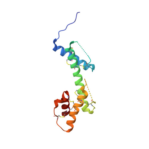
    Fibulin-2 is a multidomain, disulfide-rich, homodimeric protein which belongs to a broader extracellular matrix family. It plays an important role in the development of elastic fiber structures. Malfunction of fibulin due to mutation or poor expression can result in a variety of diseases including synpolydactyly, limb abnormalities, eye disorders leading to blindness, cardiovascular diseases and cancer. Traditionally, fibulins have either been produced in mammalian cell systems or were isolated from the extracellular matrix, a procedure that results in poor availability for structural and functional studies. Here, we produced seven fibulin-2 constructs covering 62% of the mature protein (749 out of 1195 residues) using a prokaryotic expression system. Biophysical studies confirm that the purified constructs are folded and that the presence of disulfide bonds within the constructs makes them extremely thermostable. In addition, we solved the first crystal structure for any fibulin isoform, a structure corresponding to the previously suggested three motifs related to anaphylatoxin. The structure reveals that the three anaphylatoxins moieties form a single-domain structure.


  • Organizational Affiliation
    • Faculty of Biochemistry and Molecular Medicine, University of Oulu, 90220, Oulu, Finland.

Macromolecule Content 

  • Total Structure Weight: 110.66 kDa 
  • Atom Count: 6,605 
  • Modeled Residue Count: 871 
  • Deposited Residue Count: 1,016 
  • Unique protein chains: 1

Macromolecules

Find similar proteins by:|  3D Structure
Entity ID: 1
MoleculeChains  Sequence LengthOrganismDetailsImage
Fibulin-2
A, B, C, D, E
A, B, C, D, E, F, G, H
127Mus musculusMutation(s): 0 
Gene Names: Fbln2
UniProt
Find proteins for P37889 (Mus musculus)
Explore P37889 
Go to UniProtKB:  P37889
Entity Groups
Sequence Clusters30% Identity50% Identity70% Identity90% Identity95% Identity100% Identity
UniProt GroupP37889
Sequence Annotations
Expand
Reference Sequence

Experimental Data & Validation

Experimental Data

  • Method: X-RAY DIFFRACTION
  • Resolution: 2.20 Å
  • R-Value Free:  0.297 (Depositor), 0.299 (DCC) 
  • R-Value Work:  0.243 (Depositor), 0.246 (DCC) 
  • R-Value Observed: 0.245 (Depositor) 
Space Group: P 1 21 1
Unit Cell:
Length ( Å )Angle ( ˚ )
a = 103.68α = 90
b = 34.51β = 94
c = 106.89γ = 90
Software Package:
Software NamePurpose
REFMACrefinement
xia2data reduction
xia2data scaling
PHENIXphasing

Structure Validation

View Full Validation Report


Entry History 

& Funding Information

Deposition Data


Funding OrganizationLocationGrant Number
Academy of FinlandFinland272573

Revision History  (Full details and data files)

  • Version 1.0: 2024-07-10
    Type: Initial release
  • Version 1.1: 2024-07-17
    Changes: Database references
  • Version 1.2: 2024-11-13
    Changes: Structure summary