Discovery and binding mode of small molecule inhibitors of the apo form of human TDO2.
Lotz-Jenne, C., Lange, R., Cren, S., Bourquin, G., Goglia, L., Kimmerlin, T., Wicki, M., Muller, M., Artico, N., Ackerknecht, S., Pfaff, P., Joesch, C., Mac Sweeney, A.(2024) Sci Rep 14: 27937-27937
- PubMed: 39537789 Search on PubMedSearch on PubMed Central
- DOI: https://doi.org/10.1038/s41598-024-78981-4
- Primary Citation Related Structures:
8QV7, 8R5Q, 8R5R, 9EZJ - PubMed Abstract:
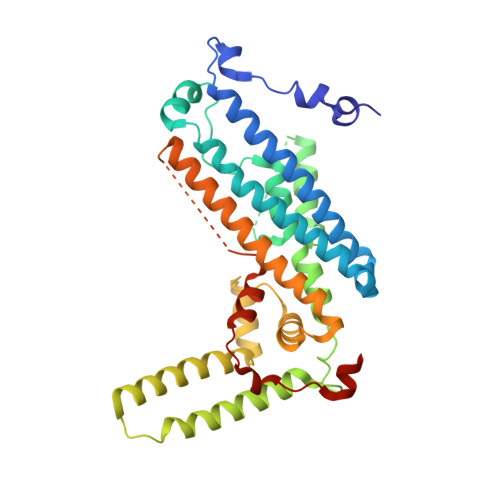
Tryptophan-2,3-dioxygenase (TDO2) and indoleamine-2,3-dioxygenase (IDO1) are structurally distinct heme enzymes that catalyze the conversion of L-tryptophan to N-formyl-kynurenine, and play important roles in metabolism, inflammation, and tumor immune surveillance. The enzymes can adopt an inactive, heme-free (apo) state or an active, heme-containing (holo) state, with the balance between them varying dynamically according to biological conditions. Inhibitors of holo-TDO2 are known but, despite several advantages of the heme-free state as a drug target, no inhibitors of apo-TDO2 have been reported. We describe the discovery of the first apo-TDO2 binding inhibitors, to our knowledge, and their inhibition of cellular TDO2 activity at low nanomolar concentrations. The crystal structure of a potent, small molecule inhibitor bound to apo-TDO2 reveals its detailed binding interactions within the large, hydrophobic heme binding pocket of the active site.
- Drug discovery, Idorsia Pharmaceuticals Limited, Hegenheimermattweg 91, Allschwil, Basel-Land, 4123, Switzerland. carina.lotz@idorsia.com.
Organizational Affiliation: