8R17 | pdb_00008r17

Crystal structure of Neurospora crassa NADase with modified C-terminus


Experimental Data Snapshot

  • Method: X-RAY DIFFRACTION
  • Resolution: 2.30 Å
  • R-Value Free: 
    0.258 (Depositor), 0.257 (DCC) 
  • R-Value Work: 
    0.234 (Depositor), 0.234 (DCC) 
  • R-Value Observed: 
    0.235 (Depositor) 

Starting Model: experimental
View more details

wwPDB Validation 3D Report Full Report

Validation slider image for 8R17

Ligand Structure Quality Assessment 


This is version 1.1 of the entry. See complete history

Literature

Evolution of fungal tuberculosis necrotizing toxin (TNT) domain-containing enzymes reveals divergent adaptations to enhance NAD cleavage.

Ferrario, E.Kallio, J.P.Emdadi, M.Stromland, O.Rack, J.G.M.Ziegler, M.

(2024) Protein Sci 33: e5071-e5071

  • DOI: https://doi.org/10.1002/pro.5071
  • Primary Citation Related Structures: 
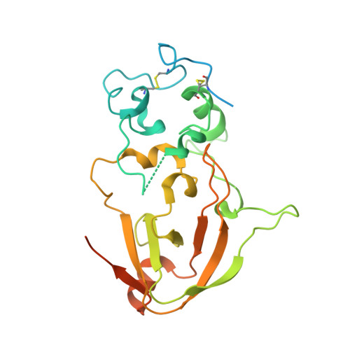
    8R15, 8R17

  • PubMed Abstract: 

    Tuberculosis necrotizing toxin (TNT) is a protein domain discovered on the outer membrane of Mycobacterium tuberculosis (Mtb), and the fungal pathogen Aspergillus fumigatus. TNT domains have pure NAD(P) hydrolytic activity, setting them apart from other NAD-cleaving domains such as ADP-ribosyl cyclase and Toll/interleukin-1 receptor homology (TIR) domains which form a wider set of products. Importantly, the Mtb TNT domain has been shown to be involved in immune evasion via depletion of the intracellular NAD pool of macrophages. Therefore, an intriguing hypothesis is that TNT domains act as "NAD killers" in host cells facilitating pathogenesis. Here, we explore the phylogenetic distribution of TNT domains and detect their presence solely in bacteria and fungi. Within fungi, we discerned six TNT clades. In addition, X-ray crystallography and AlphaFold2 modeling unveiled clade-specific strategies to promote homodimer stabilization of the fungal enzymes, namely, Ca 2+ binding, disulfide bonds, or hydrogen bonds. We show that dimer stabilization is a requirement for NADase activity and that the group-specific strategies affect the active site conformation, thereby modulating enzyme activity. Together, these findings reveal the evolutionary lineage of fungal TNT enzymes, corroborating the hypothesis of them being pure extracellular NAD (eNAD) cleavers, with possible involvement in microbial warfare and host immune evasion.


  • Organizational Affiliation
    • Department of Biomedicine, University of Bergen, Bergen, Norway.

Macromolecule Content 

  • Total Structure Weight: 29.23 kDa 
  • Atom Count: 1,554 
  • Modeled Residue Count: 188 
  • Deposited Residue Count: 259 
  • Unique protein chains: 1

Macromolecules

Find similar proteins by:|  3D Structure
Entity ID: 1
MoleculeChains  Sequence LengthOrganismDetailsImage
Conidial surface nicotinamide adenine dinucleotide glycohydrolase,Conidial surface nicotinamide adenine dinucleotide glycohydrolase nadA259Neurospora crassaAspergillus fumigatus Af293
This entity is chimeric
Mutation(s): 0 
Gene Names: NCU07948nadAAFUA_6G14470
EC: 3.2.2.5 (PDB Primary Data), 3.2.2 (PDB Primary Data)
UniProt
Find proteins for Q4WL81 (Aspergillus fumigatus (strain ATCC MYA-4609 / CBS 101355 / FGSC A1100 / Af293))
Explore Q4WL81 
Go to UniProtKB:  Q4WL81
Find proteins for Q7S936 (Neurospora crassa (strain ATCC 24698 / 74-OR23-1A / CBS 708.71 / DSM 1257 / FGSC 987))
Explore Q7S936 
Go to UniProtKB:  Q7S936
Entity Groups
Sequence Clusters30% Identity50% Identity70% Identity90% Identity95% Identity100% Identity
UniProt GroupsQ7S936Q4WL81
Glycosylation
Glycosylation Sites: 3
Sequence Annotations
Expand
Reference Sequence

Oligosaccharides

Help  
Entity ID: 2
MoleculeChains Length2D Diagram GlycosylationD Interactions
2-acetamido-2-deoxy-beta-D-glucopyranose-(1-4)-2-acetamido-2-deoxy-beta-D-glucopyranose
B
2N-Glycosylation
Glycosylation Resources
GlyTouCan: G42666HT
GlyCosmos: G42666HT
GlyGen: G42666HT

Experimental Data & Validation

Experimental Data

  • Method: X-RAY DIFFRACTION
  • Resolution: 2.30 Å
  • R-Value Free:  0.258 (Depositor), 0.257 (DCC) 
  • R-Value Work:  0.234 (Depositor), 0.234 (DCC) 
  • R-Value Observed: 0.235 (Depositor) 
Space Group: P 43 21 2
Unit Cell:
Length ( Å )Angle ( ˚ )
a = 83.756α = 90
b = 83.756β = 90
c = 103.22γ = 90
Software Package:
Software NamePurpose
PHENIXrefinement
XDSdata reduction
Aimlessdata scaling
PHASERphasing

Structure Validation

View Full Validation Report


Ligand Structure Quality Assessment 


Entry History 

& Funding Information

Deposition Data


Funding OrganizationLocationGrant Number
Norwegian Research CouncilNorway302314

Revision History  (Full details and data files)

  • Version 1.0: 2024-07-03
    Type: Initial release
  • Version 1.1: 2024-11-13
    Changes: Structure summary