8QZ7 | pdb_00008qz7

Structure of human ceramide synthase 6 (CerS6) in complex with N-palmitoyl fumonisin B1


Experimental Data Snapshot

  • Method: ELECTRON MICROSCOPY
  • Resolution: 3.00 Å
  • Aggregation State: PARTICLE 
  • Reconstruction Method: SINGLE PARTICLE 

Starting Model: in silico
View more details

wwPDB Validation 3D Report Full Report

Validation slider image for 8QZ7

This is version 1.2 of the entry. See complete history

Literature

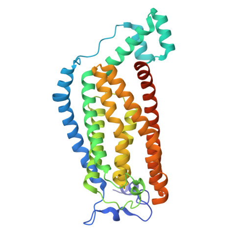
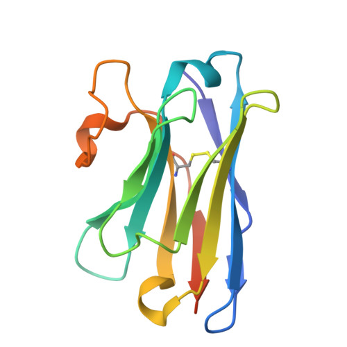
Structural basis of the mechanism and inhibition of a human ceramide synthase.

Pascoa, T.C.Pike, A.C.W.Tautermann, C.S.Chi, G.Traub, M.Quigley, A.Chalk, R.Stefanic, S.Thamm, S.Pautsch, A.Carpenter, E.P.Schnapp, G.Sauer, D.B.

(2025) Nat Struct Mol Biol 32: 431-440

  • DOI: https://doi.org/10.1038/s41594-024-01414-3
  • Primary Citation Related Structures: 
    8QZ6, 8QZ7, 9EOT

  • PubMed Abstract: 

    Ceramides are bioactive sphingolipids crucial for regulating cellular metabolism. Ceramides and dihydroceramides are synthesized by six ceramide synthase (CerS) enzymes, each with specificity for different acyl-CoA substrates. Ceramide with a 16-carbon acyl chain (C16 ceramide) has been implicated in obesity, insulin resistance and liver disease and the C16 ceramide-synthesizing CerS6 is regarded as an attractive drug target for obesity-associated disease. Despite their importance, the molecular mechanism underlying ceramide synthesis by CerS enzymes remains poorly understood. Here we report cryo-electron microscopy structures of human CerS6, capturing covalent intermediate and product-bound states. These structures, along with biochemical characterization, reveal that CerS catalysis proceeds through a ping-pong reaction mechanism involving a covalent acyl-enzyme intermediate. Notably, the product-bound structure was obtained upon reaction with the mycotoxin fumonisin B1, yielding insights into its inhibition of CerS. These results provide a framework for understanding CerS function, selectivity and inhibition and open routes for future drug discovery.


  • Organizational Affiliation
    • Centre for Medicines Discovery, Nuffield Department of Medicine, University of Oxford, Oxford, UK. tomas.pascoa@cmd.ox.ac.uk.

Macromolecule Content 

  • Total Structure Weight: 118.92 kDa 
  • Atom Count: 7,366 
  • Modeled Residue Count: 914 
  • Deposited Residue Count: 986 
  • Unique protein chains: 2

Macromolecules

Find similar proteins by:|  3D Structure
Entity ID: 1
MoleculeChains  Sequence LengthOrganismDetailsImage
Isoform 2 of Ceramide synthase 6A,
D [auth C]
357Homo sapiensMutation(s): 0 
Gene Names: CERS6LASS6
EC: 2.3.1.291 (PDB Primary Data), 2.3.1 (UniProt)
Membrane Entity: Yes 
UniProt & NIH Common Fund Data Resources
Find proteins for Q6ZMG9 (Homo sapiens)
Explore Q6ZMG9 
Go to UniProtKB:  Q6ZMG9
PHAROS:  Q6ZMG9
GTEx:  ENSG00000172292 
Entity Groups
Sequence Clusters30% Identity50% Identity70% Identity90% Identity95% Identity100% Identity
UniProt GroupQ6ZMG9
Glycosylation
Glycosylation Sites: 1Go to GlyGen: Q6ZMG9-1
Sequence Annotations
Expand
Reference Sequence
Find similar proteins by:|  3D Structure
Entity ID: 2
MoleculeChains  Sequence LengthOrganismDetailsImage
Nanobody-22B,
C [auth D]
136Vicugna pacosMutation(s): 0 

Small Molecules

Ligands 3 Unique
IDChains Name / Formula / InChI Key2D Diagram3D Interactions
XBX
(Subject of Investigation/LOI)

Query on XBX


Download:Ideal Coordinates CCD File
E [auth A],
H [auth C]
(2~{R})-2-[2-[(5~{R},6~{R},7~{S},9~{S},11~{R},16~{R},18~{S},19~{S})-6-[(3~{R})-3-carboxy-5-oxidanyl-5-oxidanylidene-pentanoyl]oxy-19-(hexadecanoylamino)-5,9-dimethyl-11,16,18-tris(oxidanyl)icosan-7-yl]oxy-2-oxidanylidene-ethyl]butanedioic acid
C50 H89 N O16
JETFIMCRGHHXJM-UHFFFAOYSA-N
PC1

Query on PC1


Download:Ideal Coordinates CCD File
F [auth A],
I [auth C]
1,2-DIACYL-SN-GLYCERO-3-PHOSPHOCHOLINE
C44 H88 N O8 P
NRJAVPSFFCBXDT-HUESYALOSA-N
NAG

Query on NAG


Download:Ideal Coordinates CCD File
G [auth A],
J [auth C]
2-acetamido-2-deoxy-beta-D-glucopyranose
C8 H15 N O6
OVRNDRQMDRJTHS-FMDGEEDCSA-N

Experimental Data & Validation

Experimental Data

  • Method: ELECTRON MICROSCOPY
  • Resolution: 3.00 Å
  • Aggregation State: PARTICLE 
  • Reconstruction Method: SINGLE PARTICLE 
EM Software:
TaskSoftware PackageVersion
MODEL REFINEMENTPHENIX1.20.1-4487
RECONSTRUCTIONcryoSPARC3.3.1

Structure Validation

View Full Validation Report


Entry History 

& Funding Information

Deposition Data


Funding OrganizationLocationGrant Number
Wellcome TrustUnited Kingdom102164/B/15/Z
Wellcome TrustUnited Kingdom106169/Z/14/Z

Revision History  (Full details and data files)

  • Version 1.0: 2024-11-13
    Type: Initial release
  • Version 1.1: 2024-11-27
    Changes: Data collection, Database references
  • Version 1.2: 2025-03-26
    Changes: Data collection, Database references