Primary Citation Related Structures: 8QSU, 8QSV, 8QSW
PubMed Abstract:
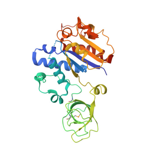
SPOUT1/CENP-32 encodes a putative SPOUT RNA methyltransferase previously identified as a mitotic chromosome associated protein. SPOUT1/CENP-32 depletion leads to centrosome detachment from the spindle poles and chromosome misalignment. Aided by gene matching platforms, here we identify 28 individuals with neurodevelopmental delays from 21 families with bi-allelic variants in SPOUT1/CENP-32 detected by exome/genome sequencing. Zebrafish spout1/cenp-32 mutants show reduction in larval head size with concomitant apoptosis likely associated with altered cell cycle progression. In vivo complementation assays in zebrafish indicate that SPOUT1/CENP-32 missense variants identified in humans are pathogenic. Crystal structure analysis of SPOUT1/CENP-32 reveals that most disease-associated missense variants are located within the catalytic domain. Additionally, SPOUT1/CENP-32 recurrent missense variants show reduced methyltransferase activity in vitro and compromised centrosome tethering to the spindle poles in human cells. Thus, SPOUT1/CENP-32 pathogenic variants cause an autosomal recessive neurodevelopmental disorder: SpADMiSS (SPOUT1 Associated Development delay Microcephaly Seizures Short stature) underpinned by mitotic spindle organization defects and consequent chromosome segregation errors.
Organizational Affiliation:
Department of Pathology and Laboratory Medicine, Children's Hospital Los Angeles, Los Angeles, CA, 90027, USA.
Keck School of Medicine, University of Southern California, Los Angeles, CA, 90033, USA.
Institute of Cell Biology, University of Edinburgh, Edinburgh, United Kingdom.
Stanley Manne Children's Research Institute, Ann & Robert H. Lurie Children's Hospital of Chicago, Chicago, IL, 60611, USA.
Departments of Pediatrics and Cell and Developmental Biology, Feinberg School of Medicine, Northwestern University, Chicago, IL, 60611, USA.
Human Molecular Genetics Lab, Health Biotechnology Division, National Institute for Biotechnology and Genetic Engineering (NIBGE-C), Faisalabad, Pakistan.
Pakistan Institute of Engineering and Applied Sciences (PIEAS), Islamabad, Pakistan.
Department of Neuromuscular Diseases, University College London, Queen Square, Institute of Neurology, WC1N 3BG, London, UK.
Department of Neurology, Vagelos College of Physicians and Surgeons, Columbia University, New York, NY, 10032, USA.
Translational Research Center for the Nervous System, Brain Cognition and Brain Disease Institute (BCBDI), Shenzhen Institute of Advanced Technology, Chinese Academy of Sciences, 518055, Shenzhen, Guangdong, China.
Faculty of Life and Health sciences, Shenzhen University of Advanced Technology, 518055, Shenzhen, Guangdong, China.
Department of Pediatrics, Chinese PLA General Hospital, Medical School of Chinese People's Liberation Army, 100853, Beijing, China.
Department of Pediatrics, Fujian Medical University Union Hospital, 350001, Fuzhou, China.
Edinburgh Protein Production Facility (EPPF), University of Edinburgh, King's Buildings, Max Born Crescent, Edinburgh, EH9 3BF, UK.
Institute of Neuroscience, Laboratory of Genetics of Brain Development, National Research Lobachevsky State University of Nizhny Novgorod, 603022, 23 Gagarin avenue, Nizhny, Novgorod, Russia.
Institute of Cell and Neurobiology, Charité Universitätsmedizin Berlin, 10117, Berlin, Charitéplatz 1, Germany.
Personalized Care (PCARE) Program, Department of Pathology and Laboratory Medicine; The Saban Research Institute, Children's Hospital Los Angeles, Los Angeles, CA, 90027, USA.
Medical Genetics, DMG Children's Rehabilitative Services, Phoenix, AZ, 85013, USA.
Division of Clinical Genetics, Department of Pediatrics, Columbia University, Vagelos College of Physicians and Surgeons, New York, NY, USA.
Institute of Human Genetics, University of Leipzig Medical Center, Leipzig, Germany.
CENTOGENE GmbH, Am Strande 7, 18055, Rostock, Germany.
Department of Development Pediatrics, The Children's Hospital and The Institute of Child Health, Multan, Pakistan.
Texas Child Neurology, Plano, TX, 75024, USA.
Neurology Consultants of Dallas, Dallas, TX, 75243, USA.
Departments of Pathology and Genetic Medicine, Johns Hopkins University School of Medicine, Baltimore, MD, 21205, USA.
Department of Paediatric Neurology, Hospital Vall d'Hebron, Barcelona, Spain.
Vall d'Hebron Research Institute, Barcelona, Spain.
Department of Paediatrics, Universitat Autònoma de Barcelona, Barcelona, Spain.
Molecular Biology CORE, Biomedical Diagnostic Center (CDB), Hospital, l Clínic de Barcelona, Barcelona, Spain.
Department of Paediatric Neurology, Hospital Infantil Niño Jesús, Madrid, Spain.
Centro de Investigación Biomédica en Red de Enfermedades Raras (CIBERER (GCV23/ER/3)), ISCIII, Madrid, Spain.
Gene Center, Department of Biochemistry, Ludwig-Maximilians Universität, Munich, Germany.
Department of Pathology and Cell Biology, Columbia University Irving Medical Center, New York, NY, 10032, USA.
Institute for Genetic Medicine Pathophysiology, Hokkaido University, Sapporo, Japan.
Department of Pediatrics, Baylor College of Medicine, Houston, TX, USA.
Stanley Manne Children's Research Institute, Ann & Robert H. Lurie Children's Hospital of Chicago, Chicago, IL, 60611, USA. eridavis@luriechildrens.org.
Departments of Pediatrics and Cell and Developmental Biology, Feinberg School of Medicine, Northwestern University, Chicago, IL, 60611, USA. eridavis@luriechildrens.org.
Institute of Cell Biology, University of Edinburgh, Edinburgh, United Kingdom. jeyaprakash.arulanandam@ed.ac.uk.
Molecular Biology CORE, Biomedical Diagnostic Center (CDB), Hospital, l Clínic de Barcelona, Barcelona, Spain. jeyaprakash.arulanandam@ed.ac.uk.
Department of Pathology and Cell Biology, Columbia University Irving Medical Center, New York, NY, 10032, USA. jl5098@cumc.columbia.edu.