8QQK

Cryo-EM structure of E. coli cytochrome bo3 quinol oxidase assembled in peptidiscs


Experimental Data Snapshot

  • Method: ELECTRON MICROSCOPY
  • Resolution: 2.80 Å
  • Aggregation State: PARTICLE 
  • Reconstruction Method: SINGLE PARTICLE 

wwPDB Validation   3D Report Full Report


This is version 1.1 of the entry. See complete history


Literature

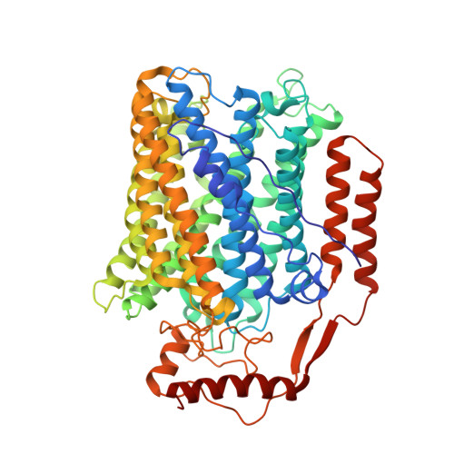
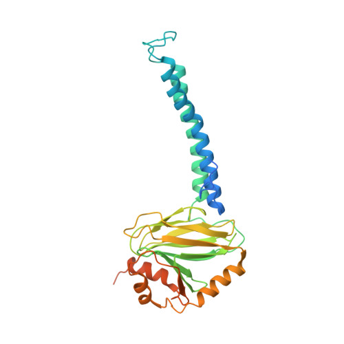
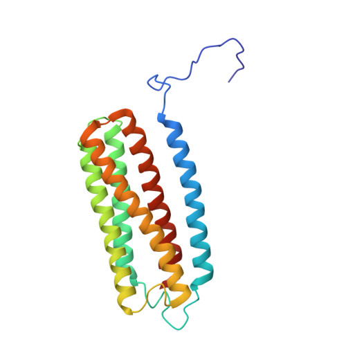
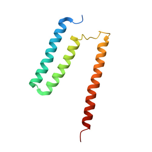
Cryo-EM structure of cytochrome bo 3 quinol oxidase assembled in peptidiscs reveals an "open" conformation for potential ubiquinone-8 release.

Gao, Y.Zhang, Y.Hakke, S.Mohren, R.Sijbers, L.J.P.M.Peters, P.J.Ravelli, R.B.G.

(2024) Biochim Biophys Acta Bioenerg 1865: 149045-149045

  • DOI: https://doi.org/10.1016/j.bbabio.2024.149045
  • Primary Citation of Related Structures:  
    8QQK

  • PubMed Abstract: 

    Cytochrome bo 3 quinol oxidase belongs to the heme‑copper-oxidoreductase (HCO) superfamily, which is part of the respiratory chain and essential for cell survival. While the reaction mechanism of cyt bo 3 has been studied extensively over the last decades, specific details about its substrate binding and product release have remained unelucidated due to the lack of structural information. Here, we report a 2.8 Å cryo-electron microscopy structure of cyt bo 3 from Escherichia coli assembled in peptidiscs. Our structural model shows a conformation for amino acids 1-41 of subunit I different from all previously published structures while the remaining parts of this enzyme are similar. Our new conformation shows a "U-shape" assembly in contrast to the transmembrane helix, named "TM0", in other reported structural models. However, TM0 blocks ubiquinone-8 (reaction product) release, suggesting that other cyt bo 3 conformations should exist. Our structural model presents experimental evidence for an "open" conformation to facilitate substrate/product exchange. This work helps further understand the reaction cycle of this oxidase, which could be a benefit for potential drug/antibiotic design for health science.


  • Organizational Affiliation

    Division of Nanoscopy, Maastricht Multimodal Molecular Imaging Institute, Maastricht University, Maastricht, the Netherlands; Division of Imaging Mass Spectrometry, Maastricht Multimodal Molecular Imaging Institute, Maastricht University, Maastricht, the Netherlands. Electronic address: y.gao@maastrichtuniversity.nl.


Macromolecules
Find similar proteins by:  (by identity cutoff)  |  3D Structure
Entity ID: 1
MoleculeChains Sequence LengthOrganismDetailsImage
Cytochrome bo(3) ubiquinol oxidase subunit 1663Escherichia coli BL21(DE3)Mutation(s): 0 
EC: 7.1.1.3
UniProt
Find proteins for P0ABI8 (Escherichia coli (strain K12))
Explore P0ABI8 
Go to UniProtKB:  P0ABI8
Entity Groups  
Sequence Clusters30% Identity50% Identity70% Identity90% Identity95% Identity100% Identity
UniProt GroupP0ABI8
Sequence Annotations
Expand
  • Reference Sequence
Find similar proteins by:  (by identity cutoff)  |  3D Structure
Entity ID: 2
MoleculeChains Sequence LengthOrganismDetailsImage
Cytochrome bo(3) ubiquinol oxidase subunit 2315Escherichia coli BL21(DE3)Mutation(s): 0 
UniProt
Find proteins for P0ABJ1 (Escherichia coli (strain K12))
Explore P0ABJ1 
Go to UniProtKB:  P0ABJ1
Entity Groups  
Sequence Clusters30% Identity50% Identity70% Identity90% Identity95% Identity100% Identity
UniProt GroupP0ABJ1
Sequence Annotations
Expand
  • Reference Sequence
Find similar proteins by:  (by identity cutoff)  |  3D Structure
Entity ID: 3
MoleculeChains Sequence LengthOrganismDetailsImage
Cytochrome bo(3) ubiquinol oxidase subunit 3204Escherichia coli BL21(DE3)Mutation(s): 0 
UniProt
Find proteins for P0ABJ3 (Escherichia coli (strain K12))
Explore P0ABJ3 
Go to UniProtKB:  P0ABJ3
Entity Groups  
Sequence Clusters30% Identity50% Identity70% Identity90% Identity95% Identity100% Identity
UniProt GroupP0ABJ3
Sequence Annotations
Expand
  • Reference Sequence
Find similar proteins by:  (by identity cutoff)  |  3D Structure
Entity ID: 4
MoleculeChains Sequence LengthOrganismDetailsImage
Cytochrome bo(3) ubiquinol oxidase subunit 4109Escherichia coli BL21(DE3)Mutation(s): 0 
UniProt
Find proteins for P0ABJ6 (Escherichia coli (strain K12))
Explore P0ABJ6 
Go to UniProtKB:  P0ABJ6
Entity Groups  
Sequence Clusters30% Identity50% Identity70% Identity90% Identity95% Identity100% Identity
UniProt GroupP0ABJ6
Sequence Annotations
Expand
  • Reference Sequence
Small Molecules
Ligands 5 Unique
IDChains Name / Formula / InChI Key2D Diagram3D Interactions
HEO
Query on HEO

Download Ideal Coordinates CCD File 
J [auth A]HEME O
C49 H58 Fe N4 O5
FRKORVCRVCLRBA-BZKSIRDQSA-L
POV
Query on POV

Download Ideal Coordinates CCD File 
E [auth A],
F [auth A],
L [auth C]
(2S)-3-(hexadecanoyloxy)-2-[(9Z)-octadec-9-enoyloxy]propyl 2-(trimethylammonio)ethyl phosphate
C42 H82 N O8 P
WTJKGGKOPKCXLL-PFDVCBLKSA-N
3PE
Query on 3PE

Download Ideal Coordinates CCD File 
G [auth A],
H [auth A],
M [auth C]
1,2-Distearoyl-sn-glycerophosphoethanolamine
C41 H82 N O8 P
LVNGJLRDBYCPGB-LDLOPFEMSA-N
HEM
Query on HEM

Download Ideal Coordinates CCD File 
I [auth A]PROTOPORPHYRIN IX CONTAINING FE
C34 H32 Fe N4 O4
KABFMIBPWCXCRK-RGGAHWMASA-L
CU
Query on CU

Download Ideal Coordinates CCD File 
K [auth A]COPPER (II) ION
Cu
JPVYNHNXODAKFH-UHFFFAOYSA-N
Experimental Data & Validation

Experimental Data

  • Method: ELECTRON MICROSCOPY
  • Resolution: 2.80 Å
  • Aggregation State: PARTICLE 
  • Reconstruction Method: SINGLE PARTICLE 
EM Software:
TaskSoftware PackageVersion
RECONSTRUCTIONRELION
MODEL REFINEMENTPHENIX

Structure Validation

View Full Validation Report


Entry History & Funding Information

Deposition Data


Funding OrganizationLocationGrant Number
Netherlands Organisation for Scientific Research (NWO)Netherlands731.016.407
Netherlands Organisation for Scientific Research (NWO)Netherlands184.034.014
Health-HollandNetherlandsLSHM21029

Revision History  (Full details and data files)

  • Version 1.0: 2024-04-24
    Type: Initial release
  • Version 1.1: 2024-05-01
    Changes: Database references