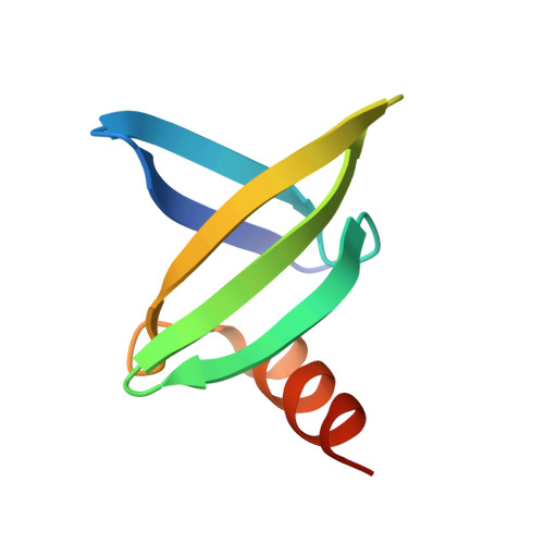
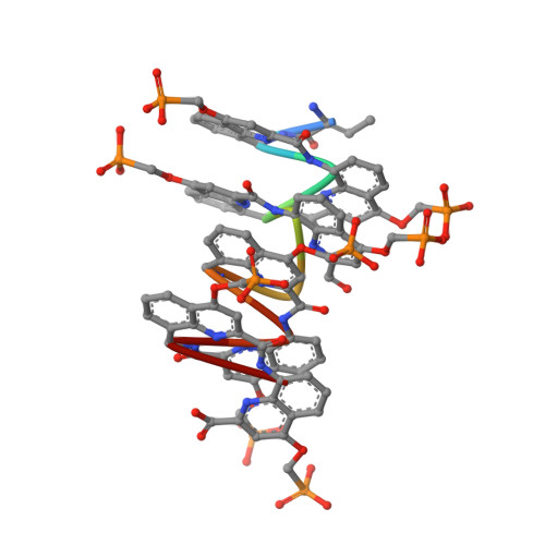
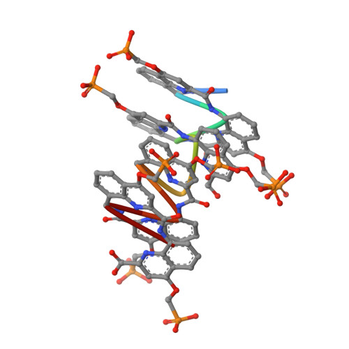
8QPC

18mer DNA mimic Foldamer with an Aromatic linker in complex with Sac7d V26A/M29A protein


Experimental Data Snapshot

  • Method: X-RAY DIFFRACTION
  • Resolution: 3.24 Å
  • R-Value Free: 0.348 
  • R-Value Work: 0.336 
  • R-Value Observed: 0.336 

wwPDB Validation   3D Report Full Report


This is version 3.0 of the entry. See complete history


Literature

DNA-Mimic Foldamer Recognition of a Chromosomal Protein

Deepak, D.Corvaglia, V.Wu, J.Allmendinger, L.Huc, I.

To be published.

Macromolecules
Find similar proteins by:  (by identity cutoff)  |  3D Structure
Entity ID: 1
MoleculeChains Sequence LengthOrganismDetailsImage
DNA-binding protein 7bA [auth AA]66Sulfolobus acidocaldariusMutation(s): 2 
Gene Names: Saci_0064
UniProt
Find proteins for P13123 (Sulfolobus acidocaldarius (strain ATCC 33909 / DSM 639 / JCM 8929 / NBRC 15157 / NCIMB 11770))
Explore P13123 
Go to UniProtKB:  P13123
Entity Groups  
Sequence Clusters30% Identity50% Identity70% Identity90% Identity95% Identity100% Identity
UniProt GroupP13123
Sequence Annotations
Expand
  • Reference Sequence

Find similar proteins by:  Sequence   |   3D Structure  

Entity ID: 2
MoleculeChains Sequence LengthOrganismDetailsImage
N-[2-(2-methyl-1,3-dioxolan-2-yl)phenyl]-2-{[5-(trifluoromethyl)pyridin-2-yl]amino}pyridine-4-carboxamide10synthetic constructMutation(s): 0 
Entity Groups  
Sequence Clusters30% Identity50% Identity70% Identity90% Identity95% Identity100% Identity
Sequence Annotations
Expand
  • Reference Sequence

Find similar proteins by:  Sequence   |   3D Structure  

Entity ID: 3
MoleculeChains Sequence LengthOrganismDetailsImage
N-[2-(2-methyl-1,3-dioxolan-2-yl)phenyl]-2-{[5-(trifluoromethyl)pyridin-2-yl]amino}pyridine-4-carboxamide10synthetic constructMutation(s): 0 
Sequence Annotations
Expand
  • Reference Sequence
Small Molecules
Modified Residues  3 Unique
IDChains TypeFormula2D DiagramParent
DBU
Query on DBU
B
PEPTIDE LINKINGC4 H7 N O2THR
V4F
Query on V4F
B
PEPTIDE LINKINGC12 H13 N2 O6 PV4F
V53
Query on V53
B
PEPTIDE LINKINGC11 H11 N2 O6 PES1
Experimental Data & Validation

Experimental Data

  • Method: X-RAY DIFFRACTION
  • Resolution: 3.24 Å
  • R-Value Free: 0.348 
  • R-Value Work: 0.336 
  • R-Value Observed: 0.336 
  • Space Group: P 64 2 2
Unit Cell:
Length ( Å )Angle ( ˚ )
a = 70.907α = 90
b = 70.907β = 90
c = 121.997γ = 120
Software Package:
Software NamePurpose
PHENIXrefinement
PHENIXrefinement
autoPROCdata reduction
Aimlessdata scaling
PHASERphasing

Structure Validation

View Full Validation Report


Entry History & Funding Information

Deposition Data


Funding OrganizationLocationGrant Number
German Research Foundation (DFG)Germany325871075 (CRC1309-C7)
European Research Council (ERC)European UnionERC-2021-ADG-320892

Revision History  (Full details and data files)

  • Version 1.0: 2023-11-08
    Type: Initial release
  • Version 2.0: 2023-11-15
    Changes: Atomic model, Data collection
  • Version 3.0: 2024-05-01
    Changes: Atomic model, Polymer sequence