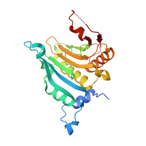
8QM4 | pdb_00008qm4

Potential drug binding sites for translation initiation factor eIF4E


Experimental Data Snapshot

  • Method: X-RAY DIFFRACTION
  • Resolution: 1.85 Å
  • R-Value Free: 
    0.217 (Depositor), 0.224 (DCC) 
  • R-Value Work: 
    0.193 (Depositor) 
  • R-Value Observed: 
    0.195 (Depositor) 

wwPDB Validation 3D Report Full Report

Validation slider image for 8QM4

Ligand Structure Quality Assessment 


This is version 1.1 of the entry. See complete history. 

Literature

Integrating fragment-based screening with targeted protein degradation and genetic rescue to explore eIF4E function.

Sharp, S.Y., Martella, M., D'Agostino, S., Milton, C.I., Ward, G., Woodhead, A.J., Richardson, C.J., Carr, M.G., Chiarparin, E., Cons, B.D., Coyle, J., East, C.E., Hiscock, S.D., Martinez-Fleites, C., Mortenson, P.N., Palmer, N., Pathuri, P., Powers, M.V., Saalau, S.M., St Denis, J.D., Swabey, K., Vinkovic, M., Walton, H., Williams, G., Clarke, P.A.

(2024) Nat Commun 15: 10037-10037

  • DOI: https://doi.org/10.1038/s41467-024-54356-1
  • Primary Citation Related Structures: 
    8QM4, 8QM5, 8QM6, 8QM7, 8QM8, 8QM9

  • PubMed Abstract: 

    Eukaryotic initiation factor 4E (eIF4E) serves as a regulatory hub for oncogene-driven protein synthesis and is considered a promising anticancer target. Here we screen a fragment library against eIF4E and identify a ligand-binding site with previously unknown function. Follow-up structure-based design yields a low nM tool compound (4, K d  = 0.09 µM; LE 0.38), which disrupts the eIF4E:eIF4G interaction, inhibits translation in cell lysates, and demonstrates target engagement with eIF4E in intact cells (EC 50  = 2 µM). By coupling targeted protein degradation with genetic rescue using eIF4E mutants, we show that disruption of both the canonical eIF4G and non-canonical binding sites is likely required to drive a strong cellular effect. This work highlights the power of fragment-based drug discovery to identify pockets in difficult-to-drug proteins and how this approach can be combined with genetic characterization and degrader technology to probe protein function in complex biological systems.


  • Organizational Affiliation: 
    • RNA Biology and Molecular Therapeutics Team, Centre for Cancer Drug Discovery, Institute of Cancer Research, London, SM2 5NG, UK.

Macromolecule Content 

  • Total Structure Weight: 50.83 kDa 
  • Atom Count: 3,484 
  • Modeled Residue Count: 402 
  • Deposited Residue Count: 430 
  • Unique protein chains: 1

Macromolecules

Find similar proteins by:|  3D Structure
Entity ID: 1
MoleculeChains  Sequence LengthOrganismDetailsImage
Eukaryotic translation initiation factor 4E
A, B
215Homo sapiensMutation(s): 0 
Gene Names: EIF4E
UniProt & NIH Common Fund Data Resources
Find proteins for P06730 (Homo sapiens)
Explore P06730 
Go to UniProtKB:  P06730
PHAROS:  P06730
GTEx:  ENSG00000151247 
Entity Groups
Sequence Clusters30% Identity50% Identity70% Identity90% Identity95% Identity100% Identity
UniProt GroupP06730
Sequence Annotations
Expand
Reference Sequence

Experimental Data & Validation

Experimental Data

  • Method: X-RAY DIFFRACTION
  • Resolution: 1.85 Å
  • R-Value Free:  0.217 (Depositor), 0.224 (DCC) 
  • R-Value Work:  0.193 (Depositor) 
  • R-Value Observed: 0.195 (Depositor) 
Space Group: P 1 21 1
Unit Cell:
Length ( Å )Angle ( ˚ )
a = 47.152α = 90
b = 69.427β = 95.92
c = 63.818γ = 90
Software Package:
Software NamePurpose
BUSTERrefinement
XDSdata reduction
SCALAdata scaling
BUSTERphasing

Structure Validation

View Full Validation Report


Ligand Structure Quality Assessment 


Entry History 

& Funding Information

Deposition Data

  • Released Date: 2025-01-15 
  • Deposition Author(s): Cleasby, A.

Funding OrganizationLocationGrant Number
Not funded--

Revision History  (Full details and data files)

  • Version 1.0: 2025-01-15
    Type: Initial release
  • Version 1.1: 2025-03-12
    Changes: Database references