8QDX

E. coli DNA gyrase bound to a DNA crossover


Experimental Data Snapshot

  • Method: ELECTRON MICROSCOPY
  • Resolution: 3.00 Å
  • Aggregation State: PARTICLE 
  • Reconstruction Method: SINGLE PARTICLE 

wwPDB Validation   3D Report Full Report


This is version 1.1 of the entry. See complete history


Literature

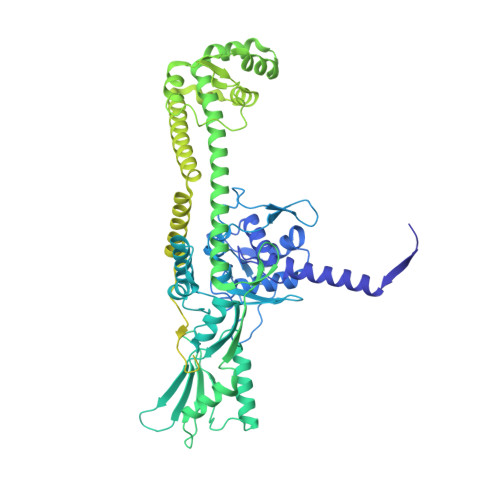
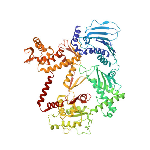
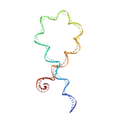
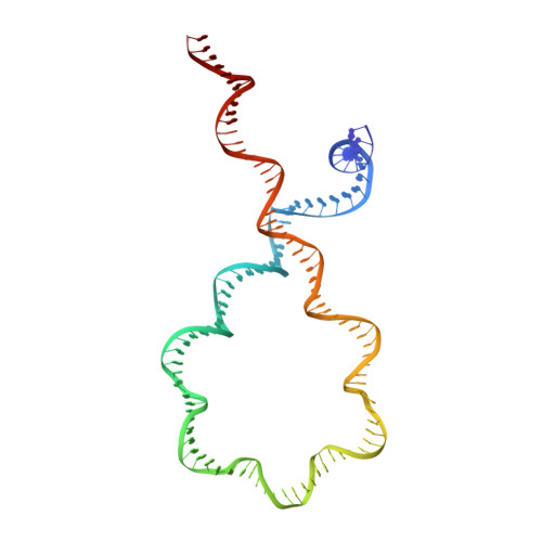
Structural basis of DNA crossover capture by Escherichia coli DNA gyrase.

Vayssieres, M.Marechal, N.Yun, L.Lopez Duran, B.Murugasamy, N.K.Fogg, J.M.Zechiedrich, L.Nadal, M.Lamour, V.

(2024) Science 384: 227-232

  • DOI: https://doi.org/10.1126/science.adl5899
  • Primary Citation of Related Structures:  
    8QDX, 8QQS, 8QQU

  • PubMed Abstract: 

    DNA supercoiling must be precisely regulated by topoisomerases to prevent DNA entanglement. The interaction of type IIA DNA topoisomerases with two DNA molecules, enabling the transport of one duplex through the transient double-stranded break of the other, remains elusive owing to structures derived solely from single linear duplex DNAs lacking topological constraints. Using cryo-electron microscopy, we solved the structure of Escherichia coli DNA gyrase bound to a negatively supercoiled minicircle DNA. We show how DNA gyrase captures a DNA crossover, revealing both conserved molecular grooves that accommodate the DNA helices. Together with molecular tweezer experiments, the structure shows that the DNA crossover is of positive chirality, reconciling the binding step of gyrase-mediated DNA relaxation and supercoiling in a single structure.


  • Organizational Affiliation

    Université de Strasbourg, Centre National de la Recherche Scientifique (CNRS), Institut national de la Recherche Médicale (INSERM), Institut de Génétique et de Biologie Moléculaire et Cellulaire (IGBMC), UMR 7104- UMR-S 1258, F-67400 Illkirch, France.


Macromolecules

Find similar proteins by:  (by identity cutoff)  |  3D Structure
Entity ID: 1
MoleculeChains Sequence LengthOrganismDetailsImage
DNA gyrase subunit A
A, C
875Escherichia coliMutation(s): 1 
Gene Names: gyrA
UniProt
Find proteins for P0AES4 (Escherichia coli (strain K12))
Explore P0AES4 
Go to UniProtKB:  P0AES4
Entity Groups  
Sequence Clusters30% Identity50% Identity70% Identity90% Identity95% Identity100% Identity
UniProt GroupP0AES4
Sequence Annotations
Expand
  • Reference Sequence
Find similar proteins by:  (by identity cutoff)  |  3D Structure
Entity ID: 2
MoleculeChains Sequence LengthOrganismDetailsImage
DNA gyrase subunit B
B, D
804Escherichia coliMutation(s): 0 
Gene Names: gyrBacrBcouhimBhisUnalCparApcbAb3699JW5625
EC: 5.6.2.2
UniProt
Find proteins for P0AES6 (Escherichia coli (strain K12))
Explore P0AES6 
Go to UniProtKB:  P0AES6
Entity Groups  
Sequence Clusters30% Identity50% Identity70% Identity90% Identity95% Identity100% Identity
UniProt GroupP0AES6
Sequence Annotations
Expand
  • Reference Sequence
Find similar nucleic acids by:  (by identity cutoff)  |  3D Structure
Entity ID: 3
MoleculeChains LengthOrganismImage
DNA minicircle118synthetic construct
Sequence Annotations
Expand
  • Reference Sequence
Find similar nucleic acids by:  (by identity cutoff)  |  3D Structure
Entity ID: 4
MoleculeChains LengthOrganismImage
DNA minicircle118synthetic construct
Sequence Annotations
Expand
  • Reference Sequence
Experimental Data & Validation

Experimental Data

  • Method: ELECTRON MICROSCOPY
  • Resolution: 3.00 Å
  • Aggregation State: PARTICLE 
  • Reconstruction Method: SINGLE PARTICLE 
EM Software:
TaskSoftware PackageVersion
MODEL REFINEMENTPHENIXv1.20.1-4487
RECONSTRUCTIONcryoSPARCV3.3.2

Structure Validation

View Full Validation Report


Entry History & Funding Information

Deposition Data


Funding OrganizationLocationGrant Number
Agence Nationale de la Recherche (ANR)France--

Revision History  (Full details and data files)

  • Version 1.0: 2024-04-10
    Type: Initial release
  • Version 1.1: 2024-04-24
    Changes: Database references