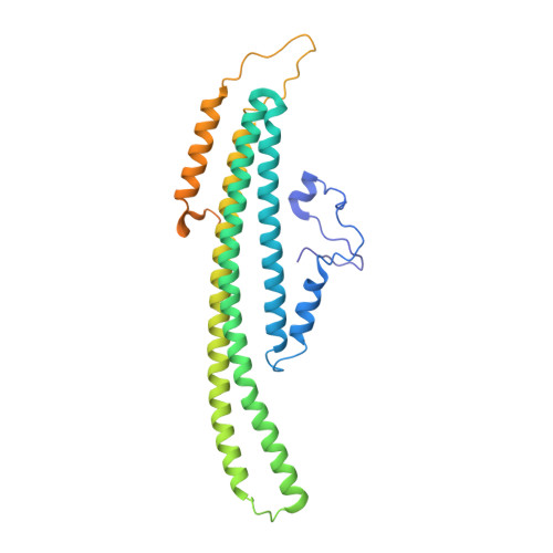
8QB7 | pdb_00008qb7

Pil1 in native eisosome lattice bound to plasma membrane microdomain


Experimental Data Snapshot

  • Method: ELECTRON MICROSCOPY
  • Resolution: 3.20 Å
  • Aggregation State: HELICAL ARRAY 
  • Reconstruction Method: SINGLE PARTICLE 

Starting Model: in silico
View more details

wwPDB Validation 3D Report Full Report

Validation slider image for 8QB7

This is version 1.4 of the entry. See complete history. 

Literature

Cryo-EM architecture of a near-native stretch-sensitive membrane microdomain.

Kefauver, J.M., Hakala, M., Zou, L., Alba, J., Espadas, J., Tettamanti, M.G., Gajic, J., Gabus, C., Campomanes, P., Estrozi, L.F., Sen, N.E., Vanni, S., Roux, A., Desfosses, A., Loewith, R.

(2024) Nature 632: 664-671

  • DOI: https://doi.org/10.1038/s41586-024-07720-6
  • Primary Citation Related Structures: 
    8QB7, 8QB8, 8QB9, 8QBB, 8QBD, 8QBE, 8QBF, 8QBG

  • PubMed Abstract: 

    Biological membranes are partitioned into functional zones termed membrane microdomains, which contain specific lipids and proteins 1-3 . The composition and organization of membrane microdomains remain controversial because few techniques are available that allow the visualization of lipids in situ without disrupting their native behaviour 3,4 . The yeast eisosome, composed of the BAR-domain proteins Pil1 and Lsp1 (hereafter, Pil1/Lsp1), scaffolds a membrane compartment that senses and responds to mechanical stress by flattening and releasing sequestered factors 5-9 . Here we isolated near-native eisosomes as helical tubules made up of a lattice of Pil1/Lsp1 bound to plasma membrane lipids, and solved their structures by helical reconstruction. Our structures reveal a striking organization of membrane lipids, and, using in vitro reconstitutions and molecular dynamics simulations, we confirmed the positioning of individual PI(4,5)P 2 , phosphatidylserine and sterol molecules sequestered beneath the Pil1/Lsp1 coat. Three-dimensional variability analysis of the native-source eisosomes revealed a dynamic stretching of the Pil1/Lsp1 lattice that affects the sequestration of these lipids. Collectively, our results support a mechanism in which stretching of the Pil1/Lsp1 lattice liberates lipids that would otherwise be anchored by the Pil1/Lsp1 coat, and thus provide mechanistic insight into how eisosome BAR-domain proteins create a mechanosensitive membrane microdomain.


  • Organizational Affiliation: 
    • Department of Molecular and Cellular Biology, University of Geneva, Geneva, Switzerland.

Macromolecule Content 

  • Total Structure Weight: 537.5 kDa 
  • Atom Count: 30,058 
  • Modeled Residue Count: 3,794 
  • Deposited Residue Count: 4,746 
  • Unique protein chains: 1

Macromolecules

Find similar proteins by:|  3D Structure
Entity ID: 1
MoleculeChains  Sequence LengthOrganismDetailsImage
Sphingolipid long chain base-responsive protein PIL1339Saccharomyces cerevisiaeMutation(s): 0 
UniProt
Find proteins for P53252 (Saccharomyces cerevisiae (strain ATCC 204508 / S288c))
Explore P53252 
Go to UniProtKB:  P53252
Entity Groups
Sequence Clusters30% Identity50% Identity70% Identity90% Identity95% Identity100% Identity
UniProt GroupP53252
Sequence Annotations
Expand
Reference Sequence

Experimental Data & Validation

Experimental Data

  • Method: ELECTRON MICROSCOPY
  • Resolution: 3.20 Å
  • Aggregation State: HELICAL ARRAY 
  • Reconstruction Method: SINGLE PARTICLE 
EM Software:
TaskSoftware PackageVersion
MODEL REFINEMENTPHENIX1.20-4459
RECONSTRUCTIONcryoSPARC4.1.2

Structure Validation

View Full Validation Report


Entry History 

& Funding Information

Deposition Data


Funding OrganizationLocationGrant Number
H2020 Marie Curie Actions of the European CommissionEuropean UnionIF-2020-101026765-MEMTOR
European Research Council (ERC)European UnionAdG TENDO
Swiss National Science FoundationSwitzerlandCRSII5_189996 METEORIC
Swiss National Science FoundationSwitzerland310030_207754

Revision History  (Full details and data files)

  • Version 1.0: 2024-07-24
    Type: Initial release
  • Version 1.1: 2024-07-31
    Changes: Data collection, Database references
  • Version 1.2: 2024-08-07
    Changes: Data collection, Database references
  • Version 1.3: 2024-08-28
    Changes: Data collection, Database references
  • Version 1.4: 2024-11-06
    Changes: Data collection, Structure summary