Structural Basis of Saccharin Derivative Inhibition of Carbonic Anhydrase IX.
Leitans, J., Kazaks, A., Bogans, J., Supuran, C.T., Akopjana, I., Ivanova, J., Zalubovskis, R., Tars, K.(2023) ChemMedChem 18: e202300454-e202300454
- PubMed: 37837260 Search on PubMed
- DOI: https://doi.org/10.1002/cmdc.202300454
- Primary Citation Related Structures:
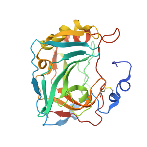
8Q18, 8Q19, 8Q1A - PubMed Abstract:
This study explores the binding mechanisms of saccharin derivatives with human carbonic anhydrase IX (hCA IX), an antitumor drug target, with the aim of facilitating the design of potent and selective inhibitors. Through the use of crystallographic analysis, we investigate the structures of hCA IX-saccharin derivative complexes, unveiling their unique binding modes that exhibit both similarities to sulfonamides and distinct orientations of the ligand tail. Our comprehensive structural insights provide information regarding the crucial interactions between the ligands and the protein, shedding light on interactions that dictate inhibitor binding and selectivity. Through a comparative analysis of the binding modes observed in hCA II and hCA IX, isoform-specific interactions are identified, offering promising strategies for the development of isoform-selective inhibitors that specifically target tumor-associated hCA IX. The findings of this study significantly deepen our understanding of the binding mechanisms of hCA inhibitors, laying a solid foundation for the rational design of more effective inhibitors.
- Latvian Biomedical Research and Study Center, Ratsupites 1, 1067, Riga, Latvia.
Organizational Affiliation: