8PWB

Crystal structure of the human METTL3-METTL14 in complex with a bisubstrate analogue (BA6)


Experimental Data Snapshot

  • Method: X-RAY DIFFRACTION
  • Resolution: 2.50 Å
  • R-Value Free: 0.244 
  • R-Value Work: 0.200 
  • R-Value Observed: 0.202 

wwPDB Validation   3D Report Full Report


Ligand Structure Quality Assessment 


This is version 1.2 of the entry. See complete history


Literature

The catalytic mechanism of the RNA methyltransferase METTL3.

Corbeski, I.Vargas-Rosales, P.A.Bedi, R.K.Deng, J.Coelho, D.Braud, E.Iannazzo, L.Li, Y.Huang, D.Etheve-Quelquejeu, M.Cui, Q.Caflisch, A.

(2024) Elife 12

  • DOI: https://doi.org/10.7554/eLife.92537
  • Primary Citation of Related Structures:  
    8PW8, 8PW9, 8PWA, 8PWB

  • PubMed Abstract: 

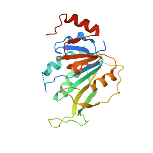
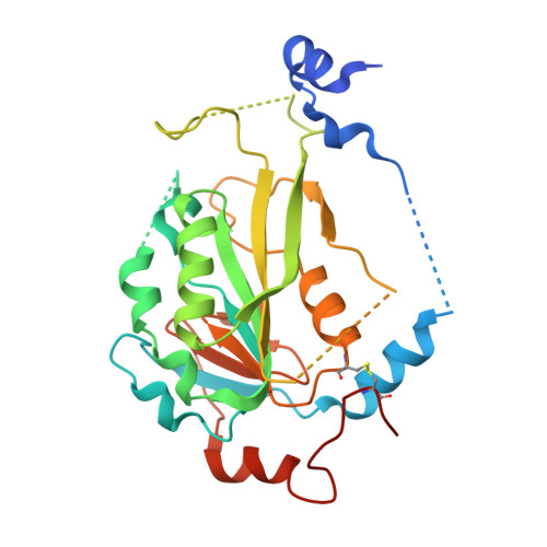
    The complex of methyltransferase-like proteins 3 and 14 (METTL3-14) is the major enzyme that deposits N 6 -methyladenosine (m 6 A) modifications on messenger RNA (mRNA) in humans. METTL3-14 plays key roles in various biological processes through its methyltransferase (MTase) activity. However, little is known about its substrate recognition and methyl transfer mechanism from its cofactor and methyl donor S -adenosylmethionine (SAM). Here, we study the MTase mechanism of METTL3-14 by a combined experimental and multiscale simulation approach using bisubstrate analogues (BAs), conjugates of a SAM-like moiety connected to the N 6 -atom of adenosine. Molecular dynamics simulations based on crystal structures of METTL3-14 with BAs suggest that the Y406 side chain of METTL3 is involved in the recruitment of adenosine and release of m 6 A. A crystal structure with a BA representing the transition state of methyl transfer shows a direct involvement of the METTL3 side chains E481 and K513 in adenosine binding which is supported by mutational analysis. Quantum mechanics/molecular mechanics (QM/MM) free energy calculations indicate that methyl transfer occurs without prior deprotonation of adenosine-N 6 . Furthermore, the QM/MM calculations provide further support for the role of electrostatic contributions of E481 and K513 to catalysis. The multidisciplinary approach used here sheds light on the (co)substrate binding mechanism, catalytic step, and (co)product release, and suggests that the latter step is rate-limiting for METTL3. The atomistic information on the substrate binding and methyl transfer reaction of METTL3 can be useful for understanding the mechanisms of other RNA MTases and for the design of transition state analogues as their inhibitors.


  • Organizational Affiliation

    Department of Biochemistry, University of Zurich, Zurich, Switzerland.


Macromolecules
Find similar proteins by:  (by identity cutoff)  |  3D Structure
Entity ID: 1
MoleculeChains Sequence LengthOrganismDetailsImage
N6-adenosine-methyltransferase catalytic subunit228Homo sapiensMutation(s): 0 
Gene Names: METTL3MTA70
EC: 2.1.1.348
UniProt & NIH Common Fund Data Resources
Find proteins for Q86U44 (Homo sapiens)
Explore Q86U44 
Go to UniProtKB:  Q86U44
PHAROS:  Q86U44
GTEx:  ENSG00000165819 
Entity Groups  
Sequence Clusters30% Identity50% Identity70% Identity90% Identity95% Identity100% Identity
UniProt GroupQ86U44
Sequence Annotations
Expand
  • Reference Sequence
Find similar proteins by:  (by identity cutoff)  |  3D Structure
Entity ID: 2
MoleculeChains Sequence LengthOrganismDetailsImage
N6-adenosine-methyltransferase non-catalytic subunit290Homo sapiensMutation(s): 0 
Gene Names: METTL14
UniProt & NIH Common Fund Data Resources
Find proteins for Q9HCE5 (Homo sapiens)
Explore Q9HCE5 
Go to UniProtKB:  Q9HCE5
PHAROS:  Q9HCE5
GTEx:  ENSG00000145388 
Entity Groups  
Sequence Clusters30% Identity50% Identity70% Identity90% Identity95% Identity100% Identity
UniProt GroupQ9HCE5
Sequence Annotations
Expand
  • Reference Sequence
Small Molecules
Ligands 2 Unique
IDChains Name / Formula / InChI Key2D Diagram3D Interactions
H3I (Subject of Investigation/LOI)
Query on H3I

Download Ideal Coordinates CCD File 
C [auth A](2~{S})-4-[[(2~{R},3~{S},4~{R},5~{R})-5-(6-aminopurin-9-yl)-3,4-bis(oxidanyl)oxolan-2-yl]methyl-(7~{H}-purin-6-ylcarbamoyl)amino]-2-azanyl-butanoic acid
C20 H24 N12 O6
OORXHLVXCSLOBF-WXSQNYJTSA-N
ACT
Query on ACT

Download Ideal Coordinates CCD File 
D [auth B]ACETATE ION
C2 H3 O2
QTBSBXVTEAMEQO-UHFFFAOYSA-M
Experimental Data & Validation

Experimental Data

  • Method: X-RAY DIFFRACTION
  • Resolution: 2.50 Å
  • R-Value Free: 0.244 
  • R-Value Work: 0.200 
  • R-Value Observed: 0.202 
  • Space Group: P 32 2 1
Unit Cell:
Length ( Å )Angle ( ˚ )
a = 63.69α = 90
b = 63.69β = 90
c = 224.77γ = 120
Software Package:
Software NamePurpose
XDSdata reduction
XSCALEdata scaling
PHASERphasing
PHENIXrefinement

Structure Validation

View Full Validation Report


Ligand Structure Quality Assessment 


Entry History & Funding Information

Deposition Data


Funding OrganizationLocationGrant Number
Swiss National Science FoundationSwitzerland310030B_189363

Revision History  (Full details and data files)

  • Version 1.0: 2023-11-08
    Type: Initial release
  • Version 1.1: 2023-12-27
    Changes: Database references
  • Version 1.2: 2024-03-27
    Changes: Database references