The Allosteric Regulation of Beta-Ureidopropionase Depends on Fine-Tuned Stability of Active-Site Loops and Subunit Interfaces.
Cederfelt, D., Badgujar, D., Au Musse, A., Lohkamp, B., Danielson, U.H., Dobritzsch, D.(2023) Biomolecules 13
- PubMed: 38136634 Search on PubMedSearch on PubMed Central
- DOI: https://doi.org/10.3390/biom13121763
- Primary Citation Related Structures:
8PT4 - PubMed Abstract:
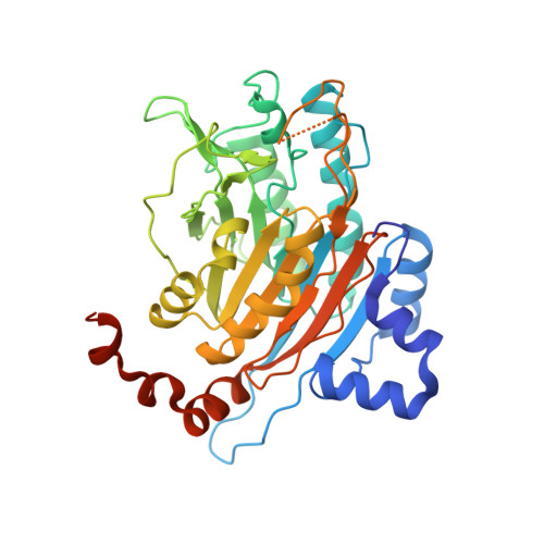
The activity of β-ureidopropionase, which catalyses the last step in the degradation of uracil, thymine, and analogous antimetabolites, is cooperatively regulated by the substrate and product of the reaction. This involves shifts in the equilibrium of the oligomeric states of the enzyme, but how these are achieved and result in changes in enzyme catalytic competence has yet to be determined. Here, the regulation of human β-ureidopropionase was further explored via site-directed mutagenesis, inhibition studies, and cryo-electron microscopy. The active-site residue E207, as well as H173 and H307 located at the dimer-dimer interface, are shown to play crucial roles in enzyme activation. Dimer association to larger assemblies requires closure of active-site loops, which positions the catalytically crucial E207 stably in the active site. H173 and H307 likely respond to ligand-induced changes in their environment with changes in their protonation states, which fine-tunes the active-site loop stability and the strength of dimer-dimer interfaces and explains the previously observed pH influence on the oligomer equilibrium. The correlation between substrate analogue structure and effect on enzyme assembly suggests that the ability to favourably interact with F205 may distinguish activators from inhibitors. The cryo-EM structure of human β-ureidopropionase assembly obtained at low pH provides first insights into the architecture of its activated state. and validates our current model of the allosteric regulation mechanism. Closed entrance loop conformations and dimer-dimer interfaces are highly conserved between human and fruit fly enzymes.
- Department of Chemistry-BMC, Uppsala University, 751 23 Uppsala, Sweden.
Organizational Affiliation: