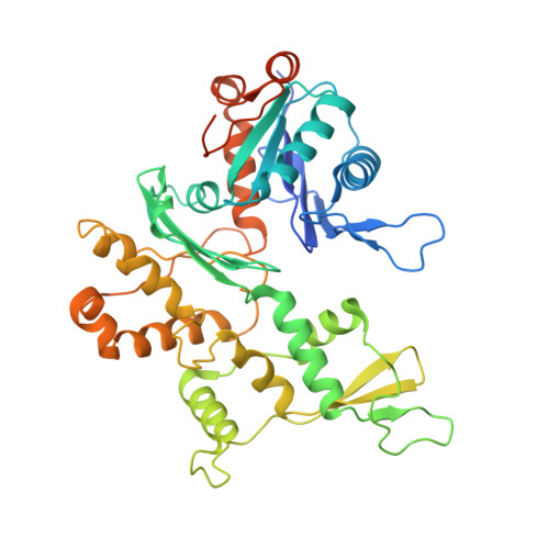
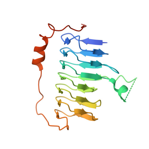
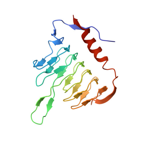
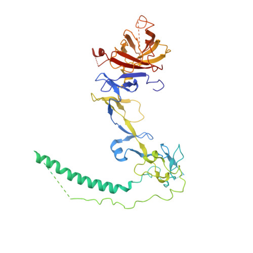
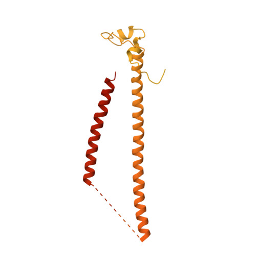
8PR4

Dynactin pointed end bound to JIP3


Experimental Data Snapshot

  • Method: ELECTRON MICROSCOPY
  • Resolution: 3.50 Å
  • Aggregation State: PARTICLE 
  • Reconstruction Method: SINGLE PARTICLE 

wwPDB Validation   3D Report Full Report


This is version 1.1 of the entry. See complete history


Literature

Molecular mechanism of dynein-dynactin complex assembly by LIS1.

Singh, K.Lau, C.K.Manigrasso, G.Gama, J.B.Gassmann, R.Carter, A.P.

(2024) Science 383: eadk8544-eadk8544

  • DOI: https://doi.org/10.1126/science.adk8544
  • Primary Citation of Related Structures:  
    8PQV, 8PQW, 8PQY, 8PQZ, 8PR0, 8PR1, 8PR2, 8PR3, 8PR4, 8PR5, 8PTK

  • PubMed Abstract: 

    Cytoplasmic dynein is a microtubule motor vital for cellular organization and division. It functions as a ~4-megadalton complex containing its cofactor dynactin and a cargo-specific coiled-coil adaptor. However, how dynein and dynactin recognize diverse adaptors, how they interact with each other during complex formation, and the role of critical regulators such as lissencephaly-1 (LIS1) protein (LIS1) remain unclear. In this study, we determined the cryo-electron microscopy structure of dynein-dynactin on microtubules with LIS1 and the lysosomal adaptor JIP3. This structure reveals the molecular basis of interactions occurring during dynein activation. We show how JIP3 activates dynein despite its atypical architecture. Unexpectedly, LIS1 binds dynactin's p150 subunit, tethering it along the length of dynein. Our data suggest that LIS1 and p150 constrain dynein-dynactin to ensure efficient complex formation.


  • Organizational Affiliation

    MRC Laboratory of Molecular Biology, Cambridge CB2 0QH, UK.


Macromolecules
Find similar proteins by:  (by identity cutoff)  |  3D Structure
Entity ID: 1
MoleculeChains Sequence LengthOrganismDetailsImage
Arp11A [auth J]417Sus scrofaMutation(s): 0 
UniProt
Find proteins for I3LHK5 (Sus scrofa)
Explore I3LHK5 
Go to UniProtKB:  I3LHK5
Entity Groups  
Sequence Clusters30% Identity50% Identity70% Identity90% Identity95% Identity100% Identity
UniProt GroupI3LHK5
Sequence Annotations
Expand
  • Reference Sequence
Find similar proteins by:  (by identity cutoff)  |  3D Structure
Entity ID: 2
MoleculeChains Sequence LengthOrganismDetailsImage
Dynactin 6B [auth U]190Sus scrofaMutation(s): 0 
UniProt
Find proteins for D0G6S1 (Sus scrofa)
Explore D0G6S1 
Go to UniProtKB:  D0G6S1
Entity Groups  
Sequence Clusters30% Identity50% Identity70% Identity90% Identity95% Identity100% Identity
UniProt GroupD0G6S1
Sequence Annotations
Expand
  • Reference Sequence
Find similar proteins by:  (by identity cutoff)  |  3D Structure
Entity ID: 3
MoleculeChains Sequence LengthOrganismDetailsImage
Dynactin subunit 5C [auth W]182Sus scrofaMutation(s): 0 
UniProt
Find proteins for A0A286ZK88 (Sus scrofa)
Explore A0A286ZK88 
Go to UniProtKB:  A0A286ZK88
Entity Groups  
Sequence Clusters30% Identity50% Identity70% Identity90% Identity95% Identity100% Identity
UniProt GroupA0A286ZK88
Sequence Annotations
Expand
  • Reference Sequence
Find similar proteins by:  (by identity cutoff)  |  3D Structure
Entity ID: 4
MoleculeChains Sequence LengthOrganismDetailsImage
Dynactin subunit 4D [auth Y]467Sus scrofaMutation(s): 0 
UniProt
Find proteins for A0A4X1TB62 (Sus scrofa)
Explore A0A4X1TB62 
Go to UniProtKB:  A0A4X1TB62
Entity Groups  
Sequence Clusters30% Identity50% Identity70% Identity90% Identity95% Identity100% Identity
UniProt GroupA0A4X1TB62
Sequence Annotations
Expand
  • Reference Sequence
Find similar proteins by:  (by identity cutoff)  |  3D Structure
Entity ID: 5
MoleculeChains Sequence LengthOrganismDetailsImage
C-Jun-amino-terminal kinase-interacting protein 3E [auth X],
F [auth x]
581Homo sapiensMutation(s): 0 
Gene Names: MAPK8IP3JIP3KIAA1066
UniProt & NIH Common Fund Data Resources
Find proteins for Q9UPT6 (Homo sapiens)
Explore Q9UPT6 
Go to UniProtKB:  Q9UPT6
PHAROS:  Q9UPT6
GTEx:  ENSG00000138834 
Entity Groups  
Sequence Clusters30% Identity50% Identity70% Identity90% Identity95% Identity100% Identity
UniProt GroupQ9UPT6
Sequence Annotations
Expand
  • Reference Sequence
Experimental Data & Validation

Experimental Data

  • Method: ELECTRON MICROSCOPY
  • Resolution: 3.50 Å
  • Aggregation State: PARTICLE 
  • Reconstruction Method: SINGLE PARTICLE 
EM Software:
TaskSoftware PackageVersion
MODEL REFINEMENTPHENIX1.20_4459:

Structure Validation

View Full Validation Report


Entry History & Funding Information

Deposition Data


Funding OrganizationLocationGrant Number
Wellcome TrustUnited Kingdom210711/Z/18/Z
Medical Research Council (MRC, United Kingdom)United KingdomMC_UP_A025_1011
European Molecular Biology Organization (EMBO)European UnionALTF 197-2021

Revision History  (Full details and data files)

  • Version 1.0: 2024-03-27
    Type: Initial release
  • Version 1.1: 2024-04-10
    Changes: Database references