8POW

Crystal Structure of the C19G variant of the membrane-bound [NiFe]-Hydrogenase from Cupriavidus necator in the air-oxidized state at 1.61 A Resolution.


Experimental Data Snapshot

  • Method: X-RAY DIFFRACTION
  • Resolution: 1.61 Å
  • R-Value Free: 0.163 
  • R-Value Work: 0.137 
  • R-Value Observed: 0.138 

wwPDB Validation   3D Report Full Report


This is version 1.0 of the entry. See complete history


Literature

Stepwise conversion of the Cys 6 [4Fe-3S] to a Cys 4 [4Fe-4S] cluster and its impact on the oxygen tolerance of [NiFe]-hydrogenase.

Schmidt, A.Kalms, J.Lorent, C.Katz, S.Frielingsdorf, S.Evans, R.M.Fritsch, J.Siebert, E.Teutloff, C.Armstrong, F.A.Zebger, I.Lenz, O.Scheerer, P.

(2023) Chem Sci 14: 11105-11120

  • DOI: https://doi.org/10.1039/d3sc03739h
  • Primary Citation of Related Structures:  
    8POU, 8POV, 8POW

  • PubMed Abstract: 

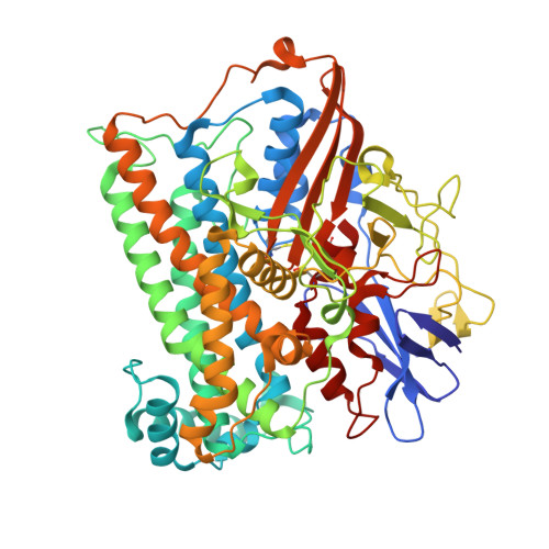
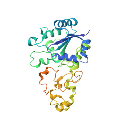
    The membrane-bound [NiFe]-hydrogenase of Cupriavidus necator is a rare example of a truly O 2 -tolerant hydrogenase. It catalyzes the oxidation of H 2 into 2e - and 2H + in the presence of high O 2 concentrations. This characteristic trait is intimately linked to the unique Cys 6 [4Fe-3S] cluster located in the proximal position to the catalytic center and coordinated by six cysteine residues. Two of these cysteines play an essential role in redox-dependent cluster plasticity, which bestows the cofactor with the capacity to mediate two redox transitions at physiological potentials. Here, we investigated the individual roles of the two additional cysteines by replacing them individually as well as simultaneously with glycine. The crystal structures of the corresponding MBH variants revealed the presence of Cys 5 [4Fe-4S] or Cys 4 [4Fe-4S] clusters of different architecture. The protein X-ray crystallography results were correlated with accompanying biochemical, spectroscopic and electrochemical data. The exchanges resulted in a diminished O 2 tolerance of all MBH variants, which was attributed to the fact that the modified proximal clusters mediated only one redox transition. The previously proposed O 2 protection mechanism that detoxifies O 2 to H 2 O using four protons and four electrons supplied by the cofactor infrastructure, is extended by our results, which suggest efficient shutdown of enzyme function by formation of a hydroxy ligand in the active site that protects the enzyme from O 2 binding under electron-deficient conditions.


  • Organizational Affiliation

    Charité - Universitätsmedizin Berlin, Corporate Member of Freie Universität Berlin and Humboldt-Universität zu Berlin, Institute of Medical Physics and Biophysics (CC2), Group Structural Biology of Cellular Signaling Charitéplatz 1 10117 Berlin Germany patrick.scheerer@charite.de.


Macromolecules
Find similar proteins by:  (by identity cutoff)  |  3D Structure
Entity ID: 1
MoleculeChains Sequence LengthOrganismDetailsImage
Uptake hydrogenase large subunitA [auth L]603Cupriavidus necator H16Mutation(s): 0 
Gene Names: hoxGPHG002
EC: 1.12.99.6
UniProt
Find proteins for P31891 (Cupriavidus necator (strain ATCC 17699 / DSM 428 / KCTC 22496 / NCIMB 10442 / H16 / Stanier 337))
Explore P31891 
Go to UniProtKB:  P31891
Entity Groups  
Sequence Clusters30% Identity50% Identity70% Identity90% Identity95% Identity100% Identity
UniProt GroupP31891
Sequence Annotations
Expand
  • Reference Sequence
Find similar proteins by:  (by identity cutoff)  |  3D Structure
Entity ID: 2
MoleculeChains Sequence LengthOrganismDetailsImage
Uptake hydrogenase small subunitB [auth S]328Cupriavidus necator H16Mutation(s): 1 
Gene Names: hoxKPHG001
EC: 1.12.99.6
UniProt
Find proteins for P31892 (Cupriavidus necator (strain ATCC 17699 / DSM 428 / KCTC 22496 / NCIMB 10442 / H16 / Stanier 337))
Explore P31892 
Go to UniProtKB:  P31892
Entity Groups  
Sequence Clusters30% Identity50% Identity70% Identity90% Identity95% Identity100% Identity
UniProt GroupP31892
Sequence Annotations
Expand
  • Reference Sequence
Small Molecules
Ligands 6 Unique
IDChains Name / Formula / InChI Key2D Diagram3D Interactions
ER2 (Subject of Investigation/LOI)
Query on ER2

Download Ideal Coordinates CCD File 
H [auth S]Fe4S4
Fe4 S4
LJBDFODJNLIPKO-UHFFFAOYSA-N
SF4
Query on SF4

Download Ideal Coordinates CCD File 
F [auth S]IRON/SULFUR CLUSTER
Fe4 S4
LJBDFODJNLIPKO-UHFFFAOYSA-N
F3S
Query on F3S

Download Ideal Coordinates CCD File 
G [auth S]FE3-S4 CLUSTER
Fe3 S4
FCXHZBQOKRZXKS-UHFFFAOYSA-N
NFV (Subject of Investigation/LOI)
Query on NFV

Download Ideal Coordinates CCD File 
C [auth L]NI-FE OXIDIZED ACTIVE CENTER
C3 Fe N2 Ni O2
MPQMGFDSXFFIQL-UHFFFAOYSA-N
CL
Query on CL

Download Ideal Coordinates CCD File 
E [auth L],
I [auth S],
J [auth S]
CHLORIDE ION
Cl
VEXZGXHMUGYJMC-UHFFFAOYSA-M
MG
Query on MG

Download Ideal Coordinates CCD File 
D [auth L]MAGNESIUM ION
Mg
JLVVSXFLKOJNIY-UHFFFAOYSA-N
Modified Residues  1 Unique
IDChains TypeFormula2D DiagramParent
CSO
Query on CSO
A [auth L]L-PEPTIDE LINKINGC3 H7 N O3 SCYS
Experimental Data & Validation

Experimental Data

  • Method: X-RAY DIFFRACTION
  • Resolution: 1.61 Å
  • R-Value Free: 0.163 
  • R-Value Work: 0.137 
  • R-Value Observed: 0.138 
  • Space Group: P 21 21 21
Unit Cell:
Length ( Å )Angle ( ˚ )
a = 72.526α = 90
b = 95.523β = 90
c = 118.868γ = 90
Software Package:
Software NamePurpose
REFMACrefinement
XDSdata reduction
SCALAdata scaling
PHASERphasing

Structure Validation

View Full Validation Report


Entry History & Funding Information

Deposition Data


Funding OrganizationLocationGrant Number
German Research Foundation (DFG)GermanyDFG Cluster of Excellence Unifying Concepts in Catalysis - Research Field D3/E3-1
German Research Foundation (DFG)GermanyUniSysCat - Germany Excellence Strategy EXC2008/1-390540038

Revision History  (Full details and data files)

  • Version 1.0: 2023-11-15
    Type: Initial release