8PEJ | pdb_00008pej

CjGH35 with a Galactosidase Activity-Based Probe


Experimental Data Snapshot

  • Method: X-RAY DIFFRACTION
  • Resolution: 1.50 Å
  • R-Value Free: 
    0.181 (Depositor), 0.180 (DCC) 
  • R-Value Work: 
    0.157 (Depositor), 0.156 (DCC) 

Starting Model: experimental
View more details

wwPDB Validation 3D Report Full Report

Validation slider image for 8PEJ

Ligand Structure Quality Assessment 


This is version 1.2 of the entry. See complete history

Literature

The development of a broad-spectrum retaining beta-exo-galactosidase activity-based probe.

Kuo, C.L.Su, Q.van den Nieuwendijk, A.M.C.H.Beenakker, T.J.M.Offen, W.A.Willems, L.I.Boot, R.G.Sarris, A.J.Marques, A.R.A.Codee, J.D.C.van der Marel, G.A.Florea, B.I.Davies, G.J.Overkleeft, H.S.Aerts, J.M.F.G.

(2023) Org Biomol Chem 21: 7813-7820

  • DOI: https://doi.org/10.1039/d3ob01261a
  • Primary Citation Related Structures: 
    8PEJ

  • PubMed Abstract: 

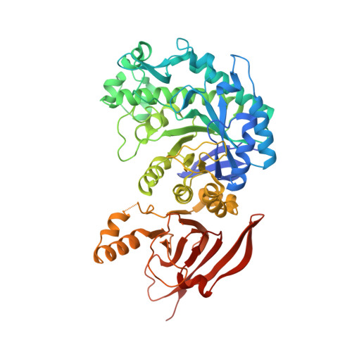
    Acid β-galactosidase (GLB1) and galactocerebrosidase (GALC) are retaining exo-β-galactosidases involved in lysosomal glycoconjugate metabolism. Deficiency of GLB1 may result in the lysosomal storage disorders GM1 gangliosidosis, Morquio B syndrome, and galactosialidosis, and deficiency of GALC may result in Krabbe disease. Activity-based protein profiling (ABPP) is a powerful technique to assess the activity of retaining glycosidases in relation to health and disease. This work describes the use of fluorescent and biotin-carrying activity-based probes (ABPs) to assess the activity of both GLB1 and GALC in cell lysates, culture media, and tissue extracts. The reported ABPs, which complement the growing list of retaining glycosidase ABPs based on configurational isomers of cyclophellitol, should assist in fundamental and clinical research on various β-galactosidases, whose inherited deficiencies cause debilitating lysosomal storage disorders.


  • Organizational Affiliation
    • Department of Medical Biochemistry, Leiden Institute of Chemistry, Leiden University, P. O. Box 9502, 2300 RA Leiden, The Netherlands. j.m.f.g.aerts@lic.leidenuniv.nl.

Macromolecule Content 

  • Total Structure Weight: 500.2 kDa 
  • Atom Count: 39,004 
  • Modeled Residue Count: 4,310 
  • Deposited Residue Count: 4,400 
  • Unique protein chains: 1

Macromolecules

Find similar proteins by:|  3D Structure
Entity ID: 1
MoleculeChains  Sequence LengthOrganismDetailsImage
Beta-galactosidase, putative, bgl35A
A, B, C, D, E
A, B, C, D, E, F, G, H
550Cellvibrio japonicus Ueda107Mutation(s): 0 
Gene Names: bgl35ACJA_2707
EC: 3.2.1.23
UniProt
Find proteins for B3PBE0 (Cellvibrio japonicus (strain Ueda107))
Explore B3PBE0 
Go to UniProtKB:  B3PBE0
Entity Groups
Sequence Clusters30% Identity50% Identity70% Identity90% Identity95% Identity100% Identity
UniProt GroupB3PBE0
Sequence Annotations
Expand
Reference Sequence

Small Molecules

Ligands 3 Unique
IDChains Name / Formula / InChI Key2D Diagram3D Interactions
YGX
(Subject of Investigation/LOI)

Query on YGX


Download:Ideal Coordinates CCD File
AA [auth D]
GB [auth H]
IA [auth E]
J [auth A]
P [auth B]
AA [auth D],
GB [auth H],
IA [auth E],
J [auth A],
P [auth B],
RA [auth F],
T [auth C],
XA [auth G]
(1~{S},2~{S},4~{S},5~{R})-6-(hydroxymethyl)cyclohexane-1,2,3,4,5-pentol
C7 H14 O6
QFYQIWDMMSKNFF-OUIVRQFQSA-N
ACT

Query on ACT


Download:Ideal Coordinates CCD File
AB [auth G]
BA [auth D]
BB [auth G]
CA [auth D]
HB [auth H]
AB [auth G],
BA [auth D],
BB [auth G],
CA [auth D],
HB [auth H],
IB [auth H],
JA [auth E],
K [auth A],
KA [auth E],
SA [auth F],
TA [auth F],
YA [auth G],
Z [auth D],
ZA [auth G]
ACETATE ION
C2 H3 O2
QTBSBXVTEAMEQO-UHFFFAOYSA-M
NA

Query on NA


Download:Ideal Coordinates CCD File
CB [auth G]
DA [auth D]
DB [auth G]
EA [auth D]
EB [auth G]
CB [auth G],
DA [auth D],
DB [auth G],
EA [auth D],
EB [auth G],
FA [auth E],
FB [auth H],
GA [auth E],
HA [auth E],
I [auth A],
JB [auth H],
KB [auth H],
L [auth A],
LA [auth E],
LB [auth H],
M [auth A],
MA [auth E],
MB [auth H],
N [auth A],
NA [auth E],
O [auth B],
OA [auth F],
PA [auth F],
Q [auth B],
QA [auth F],
R [auth C],
S [auth C],
U [auth C],
UA [auth F],
V [auth C],
VA [auth G],
W [auth D],
WA [auth G],
X [auth D],
Y [auth D]
SODIUM ION
Na
FKNQFGJONOIPTF-UHFFFAOYSA-N

Experimental Data & Validation

Experimental Data

  • Method: X-RAY DIFFRACTION
  • Resolution: 1.50 Å
  • R-Value Free:  0.181 (Depositor), 0.180 (DCC) 
  • R-Value Work:  0.157 (Depositor), 0.156 (DCC) 
Space Group: P 1
Unit Cell:
Length ( Å )Angle ( ˚ )
a = 99.316α = 90.236
b = 116.212β = 89.889
c = 116.396γ = 90.094
Software Package:
Software NamePurpose
REFMACrefinement
REFMACrefinement
Aimlessdata scaling
REFMACphasing

Structure Validation

View Full Validation Report


Ligand Structure Quality Assessment 


Entry History 

& Funding Information

Deposition Data


Funding OrganizationLocationGrant Number
Biotechnology and Biological Sciences Research Council (BBSRC)United KingdomBB/R001162/1
European Research Council (ERC)European Union951231

Revision History  (Full details and data files)

  • Version 1.0: 2023-10-04
    Type: Initial release
  • Version 1.1: 2023-10-18
    Changes: Database references
  • Version 1.2: 2024-10-23
    Changes: Structure summary