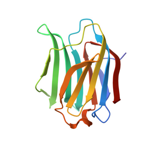
8PBF

Galectin-3C in complex with a triazolesulfane derivative


Experimental Data Snapshot

  • Method: X-RAY DIFFRACTION
  • Resolution: 1.14 Å
  • R-Value Free: 0.176 
  • R-Value Work: 0.149 
  • R-Value Observed: 0.150 

Starting Model: experimental
View more details

wwPDB Validation   3D Report Full Report


Ligand Structure Quality Assessment 


This is version 1.1 of the entry. See complete history


Literature

Ligand Sulfur Oxidation State Progressively Alters Galectin-3-Ligand Complex Conformations To Induce Affinity-Influencing Hydrogen Bonds.

Mahanti, M.Pal, K.B.Kumar, R.Schulze, M.Leffler, H.Logan, D.T.Nilsson, U.J.

(2023) J Med Chem 66: 14716-14723

  • DOI: https://doi.org/10.1021/acs.jmedchem.3c01223
  • Primary Citation of Related Structures:  
    8PBF, 8PF9, 8PFF

  • PubMed Abstract: 

    Galectins play biological roles in immune regulation and tumor progression. Ligands with high affinity for the shallow, hydrophilic galectin-3 ligand binding site rely primarily on a galactose core with appended aryltriazole moieties, making hydrophobic interactions and π-stacking. We designed and synthesized phenyl sulfone, sulfoxide, and sulfide-triazolyl thiogalactoside derivatives to create affinity-enhancing hydrogen bonds, hydrophobic and π-interactions. Crystal structures and thermodynamic analyses revealed that the sulfoxide and sulfone ligands form hydrogen bonds while retaining π-interactions, resulting in improved affinities and unique binding poses. The sulfoxide, bearing one hydrogen bond acceptor, leads to an affinity decrease compared to the sulfide, whereas the corresponding sulfone forms three hydrogen bonds, two directly with Asn and Arg side chains and one water-mediated to an Asp side chain, respectively, which alters the complex structure and increases affinity. These findings highlight that the sulfur oxidation state influences both the interaction thermodynamics and structure.


  • Organizational Affiliation

    Department of Chemistry, Lund University, Box 124, SE-221 00 Lund, Sweden.


Macromolecules
Find similar proteins by:  (by identity cutoff)  |  3D Structure
Entity ID: 1
MoleculeChains Sequence LengthOrganismDetailsImage
Galectin-3138Homo sapiensMutation(s): 0 
Gene Names: LGALS3MAC2
UniProt & NIH Common Fund Data Resources
Find proteins for P17931 (Homo sapiens)
Explore P17931 
Go to UniProtKB:  P17931
PHAROS:  P17931
GTEx:  ENSG00000131981 
Entity Groups  
Sequence Clusters30% Identity50% Identity70% Identity90% Identity95% Identity100% Identity
UniProt GroupP17931
Sequence Annotations
Expand
  • Reference Sequence
Small Molecules
Ligands 2 Unique
IDChains Name / Formula / InChI Key2D Diagram3D Interactions
Y58 (Subject of Investigation/LOI)
Query on Y58

Download Ideal Coordinates CCD File 
B [auth A](2~{R},3~{S},4~{S},5~{R},6~{S})-2-(hydroxymethyl)-6-[(2~{S},3~{R},4~{S},5~{R},6~{R})-6-(hydroxymethyl)-3,5-bis(oxidanyl)-4-(4-phenylsulfanyl-1,2,3-triazol-1-yl)oxan-2-yl]sulfanyl-oxane-3,4,5-triol
C20 H27 N3 O9 S2
ROQDROPGJRNKKP-VTOAZQAJSA-N
SCN
Query on SCN

Download Ideal Coordinates CCD File 
C [auth A]THIOCYANATE ION
C N S
ZMZDMBWJUHKJPS-UHFFFAOYSA-M
Experimental Data & Validation

Experimental Data

  • Method: X-RAY DIFFRACTION
  • Resolution: 1.14 Å
  • R-Value Free: 0.176 
  • R-Value Work: 0.149 
  • R-Value Observed: 0.150 
  • Space Group: P 21 21 21
Unit Cell:
Length ( Å )Angle ( ˚ )
a = 36.47α = 90
b = 57.005β = 90
c = 61.927γ = 90
Software Package:
Software NamePurpose
PHENIXrefinement
XDSdata reduction
XSCALEdata scaling

Structure Validation

View Full Validation Report


Ligand Structure Quality Assessment 


Entry History & Funding Information

Deposition Data


Funding OrganizationLocationGrant Number
Swedish Research CouncilSweden2016-04855
Swedish Research CouncilSweden2016-036767
Swedish Research CouncilSweden2020-03317
The Crafoord FoundationSweden20190803

Revision History  (Full details and data files)

  • Version 1.0: 2023-11-08
    Type: Initial release
  • Version 1.1: 2023-11-22
    Changes: Database references