8P72

Cryo-EM structure of CAK in complex with inhibitor ICEC0768


Experimental Data Snapshot

  • Method: ELECTRON MICROSCOPY
  • Resolution: 1.90 Å
  • Aggregation State: PARTICLE 
  • Reconstruction Method: SINGLE PARTICLE 

wwPDB Validation   3D Report Full Report


This is version 1.1 of the entry. See complete history


Literature

High-resolution cryo-EM of the human CDK-activating kinase for structure-based drug design.

Cushing, V.I.Koh, A.F.Feng, J.Jurgaityte, K.Bondke, A.Kroll, S.H.B.Barbazanges, M.Scheiper, B.Bahl, A.K.Barrett, A.G.M.Ali, S.Kotecha, A.Greber, B.J.

(2024) Nat Commun 15: 2265-2265

  • DOI: https://doi.org/10.1038/s41467-024-46375-9
  • Primary Citation of Related Structures:  
    8ORM, 8P6V, 8P6W, 8P6X, 8P6Y, 8P6Z, 8P70, 8P71, 8P72, 8P73, 8P74, 8P75, 8P76, 8P77, 8P78, 8P79, 8P7L, 8PLZ

  • PubMed Abstract: 

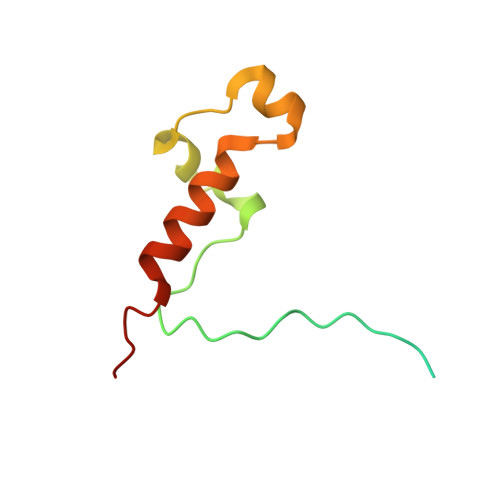
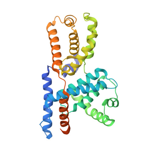
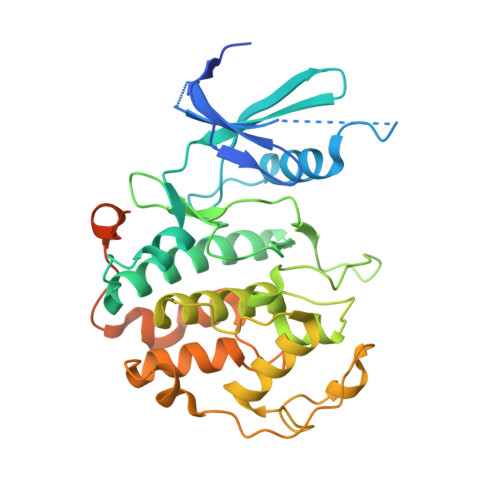
    Rational design of next-generation therapeutics can be facilitated by high-resolution structures of drug targets bound to small-molecule inhibitors. However, application of structure-based methods to macromolecules refractory to crystallization has been hampered by the often-limiting resolution and throughput of cryogenic electron microscopy (cryo-EM). Here, we use high-resolution cryo-EM to determine structures of the CDK-activating kinase, a master regulator of cell growth and division, in its free and nucleotide-bound states and in complex with 15 inhibitors at up to 1.8 Å resolution. Our structures provide detailed insight into inhibitor interactions and networks of water molecules in the active site of cyclin-dependent kinase 7 and provide insights into the mechanisms contributing to inhibitor selectivity, thereby providing the basis for rational design of next-generation therapeutics. These results establish a methodological framework for the use of high-resolution cryo-EM in structure-based drug design.


  • Organizational Affiliation

    The Institute of Cancer Research, Chester Beatty Laboratories, 237 Fulham Road, London, SW3 6JB, UK.


Macromolecules
Find similar proteins by:  (by identity cutoff)  |  3D Structure
Entity ID: 1
MoleculeChains Sequence LengthOrganismDetailsImage
CDK-activating kinase assembly factor MAT1A [auth H]93Homo sapiensMutation(s): 0 
Gene Names: MNAT1CAP35MAT1RNF66
UniProt & NIH Common Fund Data Resources
Find proteins for P51948 (Homo sapiens)
Explore P51948 
Go to UniProtKB:  P51948
PHAROS:  P51948
GTEx:  ENSG00000020426 
Entity Groups  
Sequence Clusters30% Identity50% Identity70% Identity90% Identity95% Identity100% Identity
UniProt GroupP51948
Sequence Annotations
Expand
  • Reference Sequence
Find similar proteins by:  (by identity cutoff)  |  3D Structure
Entity ID: 2
MoleculeChains Sequence LengthOrganismDetailsImage
Cyclin-HB [auth I]324Homo sapiensMutation(s): 1 
Gene Names: CCNH
UniProt & NIH Common Fund Data Resources
Find proteins for P51946 (Homo sapiens)
Explore P51946 
Go to UniProtKB:  P51946
PHAROS:  P51946
GTEx:  ENSG00000134480 
Entity Groups  
Sequence Clusters30% Identity50% Identity70% Identity90% Identity95% Identity100% Identity
UniProt GroupP51946
Sequence Annotations
Expand
  • Reference Sequence
Find similar proteins by:  (by identity cutoff)  |  3D Structure
Entity ID: 3
MoleculeChains Sequence LengthOrganismDetailsImage
Cyclin-dependent kinase 7C [auth J]349Homo sapiensMutation(s): 0 
Gene Names: CDK7CAKCAK1CDKN7MO15STK1
EC: 2.7.11.22 (PDB Primary Data), 2.7.11.23 (PDB Primary Data)
UniProt & NIH Common Fund Data Resources
Find proteins for P50613 (Homo sapiens)
Explore P50613 
Go to UniProtKB:  P50613
PHAROS:  P50613
GTEx:  ENSG00000134058 
Entity Groups  
Sequence Clusters30% Identity50% Identity70% Identity90% Identity95% Identity100% Identity
UniProt GroupP50613
Sequence Annotations
Expand
  • Reference Sequence
Small Molecules
Ligands 1 Unique
IDChains Name / Formula / InChI Key2D Diagram3D Interactions
X2Z (Subject of Investigation/LOI)
Query on X2Z

Download Ideal Coordinates CCD File 
D [auth J]~{N}7-(phenylmethyl)-~{N}5-[(3~{S})-piperidin-3-yl]-3-propan-2-yl-pyrazolo[1,5-a]pyrimidine-5,7-diamine
C21 H28 N6
HVPAERPIODSNPK-KRWDZBQOSA-N
Experimental Data & Validation

Experimental Data

  • Method: ELECTRON MICROSCOPY
  • Resolution: 1.90 Å
  • Aggregation State: PARTICLE 
  • Reconstruction Method: SINGLE PARTICLE 
EM Software:
TaskSoftware PackageVersion
MODEL REFINEMENTPHENIX1.20.1
RECONSTRUCTIONRELION4.0 beta

Structure Validation

View Full Validation Report


Entry History & Funding Information

Deposition Data


Funding OrganizationLocationGrant Number
Medical Research Council (MRC, United Kingdom)United KingdomMR/V009354/1

Revision History  (Full details and data files)

  • Version 1.0: 2024-03-20
    Type: Initial release
  • Version 1.1: 2024-03-27
    Changes: Database references