8P6I

Crystal structure of the 139H2 Fab fragment bound to Muc1 peptide epitope


Experimental Data Snapshot

  • Method: X-RAY DIFFRACTION
  • Resolution: 2.50 Å
  • R-Value Free: 0.254 
  • R-Value Work: 0.203 

wwPDB Validation   3D Report Full Report


This is version 1.1 of the entry. See complete history


Literature

Reverse-engineering the anti-MUC1 antibody 139H2 by mass spectrometry-based de novo sequencing.

Peng, W.Giesbers, K.C.Siborova, M.Beugelink, J.W.Pronker, M.F.Schulte, D.Hilkens, J.Janssen, B.J.Strijbis, K.Snijder, J.

(2024) Life Sci Alliance 7

  • DOI: https://doi.org/10.26508/lsa.202302366
  • Primary Citation of Related Structures:  
    8P6I

  • PubMed Abstract: 

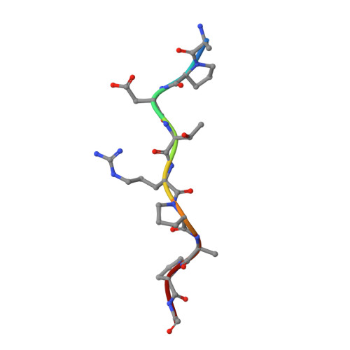
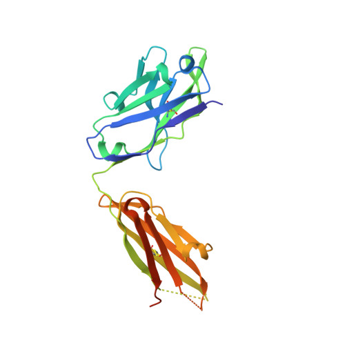
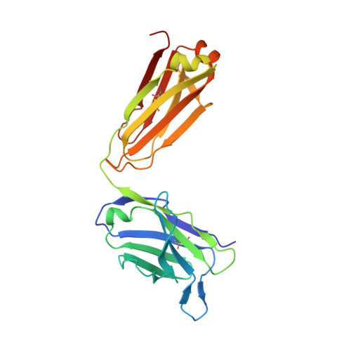
    Mucin 1 (MUC1) is a transmembrane mucin expressed at the apical surface of epithelial cells at mucosal surfaces. MUC1 has a barrier function against bacterial invasion and is well known for its aberrant expression and glycosylation in adenocarcinomas. The MUC1 extracellular domain contains a variable number of tandem repeats (VNTR) of 20 amino acids, which are heavily O -linked glycosylated. Monoclonal antibodies against the MUC1 VNTR are powerful research tools with applications in the diagnosis and treatment of MUC1-expressing cancers. Here, we report direct mass spectrometry-based sequencing of anti-MUC1 hybridoma-derived 139H2 IgG, enabling reverse-engineering of the functional recombinant monoclonal antibody. The crystal structure of the 139H2 Fab fragment in complex with the MUC1 epitope was solved, revealing the molecular basis of 139H2 binding specificity to MUC1 and its tolerance to O -glycosylation of the VNTR. The available sequence of 139H2 will allow further development of MUC1-related diagnostic, targeting, and treatment strategies.


  • Organizational Affiliation

    https://ror.org/04pp8hn57 Biomolecular Mass Spectrometry and Proteomics, Bijvoet Center for Biomolecular Research and Utrecht Institute of Pharmaceutical Sciences, Utrecht University, Utrecht, Netherlands.


Macromolecules

Find similar proteins by:  Sequence   |   3D Structure  

Entity ID: 1
MoleculeChains Sequence LengthOrganismDetailsImage
Mucin-1A,
D [auth a]
9Homo sapiensMutation(s): 0 
UniProt & NIH Common Fund Data Resources
Find proteins for P15941 (Homo sapiens)
Explore P15941 
Go to UniProtKB:  P15941
PHAROS:  P15941
GTEx:  ENSG00000185499 
Entity Groups  
UniProt GroupP15941
Sequence Annotations
Expand
  • Reference Sequence
Find similar proteins by:  (by identity cutoff)  |  3D Structure
Entity ID: 2
MoleculeChains Sequence LengthOrganismDetailsImage
139H2 HCB [auth H],
E [auth h]
236Homo sapiensMutation(s): 0 
Entity Groups  
Sequence Clusters30% Identity50% Identity70% Identity90% Identity95% Identity100% Identity
Sequence Annotations
Expand
  • Reference Sequence
Find similar proteins by:  (by identity cutoff)  |  3D Structure
Entity ID: 3
MoleculeChains Sequence LengthOrganismDetailsImage
139H2 LCC [auth L],
F [auth l]
220Homo sapiensMutation(s): 0 
Entity Groups  
Sequence Clusters30% Identity50% Identity70% Identity90% Identity95% Identity100% Identity
Sequence Annotations
Expand
  • Reference Sequence
Experimental Data & Validation

Experimental Data

  • Method: X-RAY DIFFRACTION
  • Resolution: 2.50 Å
  • R-Value Free: 0.254 
  • R-Value Work: 0.203 
  • Space Group: C 1 2 1
Unit Cell:
Length ( Å )Angle ( ˚ )
a = 211.833α = 90
b = 42.757β = 122.306
c = 129.064γ = 90
Software Package:
Software NamePurpose
REFMACrefinement
xia2.multiplexdata reduction
Aimlessdata scaling
PHASERphasing

Structure Validation

View Full Validation Report


Entry History & Funding Information

Deposition Data


Funding OrganizationLocationGrant Number
Netherlands Organisation for Scientific Research (NWO)Netherlands--

Revision History  (Full details and data files)

  • Version 1.0: 2024-04-03
    Type: Initial release
  • Version 1.1: 2024-04-10
    Changes: Refinement description