8P5V

Single particle cryo-EM structure of homohexameric 2-oxoglutarate dehydrogenase OdhA from Corynebacterium glutamicum in complex with the product succinyl-CoA


Experimental Data Snapshot

  • Method: ELECTRON MICROSCOPY
  • Resolution: 2.07 Å
  • Aggregation State: PARTICLE 
  • Reconstruction Method: SINGLE PARTICLE 

wwPDB Validation   3D Report Full Report


This is version 1.1 of the entry. See complete history


Literature

High resolution cryo-EM and crystallographic snapshots of the actinobacterial two-in-one 2-oxoglutarate dehydrogenase.

Yang, L.Wagner, T.Mechaly, A.Boyko, A.Bruch, E.M.Megrian, D.Gubellini, F.Alzari, P.M.Bellinzoni, M.

(2023) Nat Commun 14: 4851-4851

  • DOI: https://doi.org/10.1038/s41467-023-40253-6
  • Primary Citation of Related Structures:  
    8P5R, 8P5S, 8P5T, 8P5U, 8P5V, 8P5W, 8P5X

  • PubMed Abstract: 

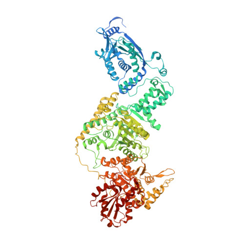
    Actinobacteria possess unique ways to regulate the oxoglutarate metabolic node. Contrary to most organisms in which three enzymes compose the 2-oxoglutarate dehydrogenase complex (ODH), actinobacteria rely on a two-in-one protein (OdhA) in which both the oxidative decarboxylation and succinyl transferase steps are carried out by the same polypeptide. Here we describe high-resolution cryo-EM and crystallographic snapshots of representative enzymes from Mycobacterium smegmatis and Corynebacterium glutamicum, showing that OdhA is an 800-kDa homohexamer that assembles into a three-blade propeller shape. The obligate trimeric and dimeric states of the acyltransferase and dehydrogenase domains, respectively, are critical for maintaining the overall assembly, where both domains interact via subtle readjustments of their interfaces. Complexes obtained with substrate analogues, reaction products and allosteric regulators illustrate how these domains operate. Furthermore, we provide additional insights into the phosphorylation-dependent regulation of this enzymatic machinery by the signalling protein OdhI.


  • Organizational Affiliation

    Institut Pasteur, Université Paris Cité, CNRS UMR3528, Unité de Microbiologie Structurale, F-75015, Paris, France.


Macromolecules
Find similar proteins by:  (by identity cutoff)  |  3D Structure
Entity ID: 1
MoleculeChains Sequence LengthOrganismDetailsImage
2-oxoglutarate dehydrogenase E1/E2 component
A, B, C, D, E
A, B, C, D, E, F
1,223Corynebacterium glutamicum ATCC 13032Mutation(s): 0 
Gene Names: odhACgl1129cg1280
EC: 1.2.4.2 (PDB Primary Data), 2.3.1.61 (PDB Primary Data)
UniProt
Find proteins for Q8NRC3 (Corynebacterium glutamicum (strain ATCC 13032 / DSM 20300 / BCRC 11384 / JCM 1318 / LMG 3730 / NCIMB 10025))
Explore Q8NRC3 
Go to UniProtKB:  Q8NRC3
Entity Groups  
Sequence Clusters30% Identity50% Identity70% Identity90% Identity95% Identity100% Identity
UniProt GroupQ8NRC3
Sequence Annotations
Expand
  • Reference Sequence
Small Molecules
Ligands 4 Unique
IDChains Name / Formula / InChI Key2D Diagram3D Interactions
SCA (Subject of Investigation/LOI)
Query on SCA

Download Ideal Coordinates CCD File 
DA [auth F]
J [auth A]
N [auth B]
R [auth C]
V [auth D]
DA [auth F],
J [auth A],
N [auth B],
R [auth C],
V [auth D],
Z [auth E]
SUCCINYL-COENZYME A
C25 H40 N7 O19 P3 S
VNOYUJKHFWYWIR-ITIYDSSPSA-N
ACO (Subject of Investigation/LOI)
Query on ACO

Download Ideal Coordinates CCD File 
BA [auth F]
H [auth A]
L [auth B]
P [auth C]
T [auth D]
BA [auth F],
H [auth A],
L [auth B],
P [auth C],
T [auth D],
X [auth E]
ACETYL COENZYME *A
C23 H38 N7 O17 P3 S
ZSLZBFCDCINBPY-ZSJPKINUSA-N
TPP (Subject of Investigation/LOI)
Query on TPP

Download Ideal Coordinates CCD File 
CA [auth F]
I [auth A]
M [auth B]
Q [auth C]
U [auth D]
CA [auth F],
I [auth A],
M [auth B],
Q [auth C],
U [auth D],
Y [auth E]
THIAMINE DIPHOSPHATE
C12 H19 N4 O7 P2 S
AYEKOFBPNLCAJY-UHFFFAOYSA-O
MG (Subject of Investigation/LOI)
Query on MG

Download Ideal Coordinates CCD File 
AA [auth F]
G [auth A]
K [auth B]
O [auth C]
S [auth D]
AA [auth F],
G [auth A],
K [auth B],
O [auth C],
S [auth D],
W [auth E]
MAGNESIUM ION
Mg
JLVVSXFLKOJNIY-UHFFFAOYSA-N
Experimental Data & Validation

Experimental Data

  • Method: ELECTRON MICROSCOPY
  • Resolution: 2.07 Å
  • Aggregation State: PARTICLE 
  • Reconstruction Method: SINGLE PARTICLE 
EM Software:
TaskSoftware PackageVersion
MODEL REFINEMENTREFMAC
RECONSTRUCTIONcryoSPARC

Structure Validation

View Full Validation Report


Entry History & Funding Information

Deposition Data


Funding OrganizationLocationGrant Number
Agence Nationale de la Recherche (ANR)FranceANR-13-JSV8-0003
Agence Nationale de la Recherche (ANR)FranceANR-18-CE92-0003

Revision History  (Full details and data files)

  • Version 1.0: 2023-08-16
    Type: Initial release
  • Version 1.1: 2023-08-23
    Changes: Database references