8P4R | pdb_00008p4r

In situ structure average of GroEL14-GroES14 complexes in Escherichia coli cytosol obtained by cryo electron tomography


Experimental Data Snapshot

  • Method: ELECTRON MICROSCOPY
  • Resolution: 11.9 Å
  • Aggregation State: CELL 
  • Reconstruction Method: SUBTOMOGRAM AVERAGING 

wwPDB Validation 3D Report Full Report

Validation slider image for 8P4R

This is version 1.3 of the entry. See complete history

Literature

Visualizing chaperonin function in situ by cryo-electron tomography.

Wagner, J.Carvajal, A.I.Bracher, A.Beck, F.Wan, W.Bohn, S.Korner, R.Baumeister, W.Fernandez-Busnadiego, R.Hartl, F.U.

(2024) Nature 633: 459-464

  • DOI: https://doi.org/10.1038/s41586-024-07843-w
  • Primary Citation Related Structures: 
    8P4M, 8P4N, 8P4O, 8P4P, 8P4R, 8QXS, 8QXT, 8QXU, 8QXV

  • PubMed Abstract: 

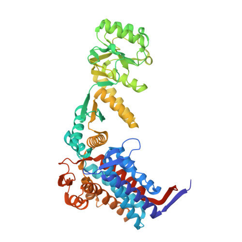
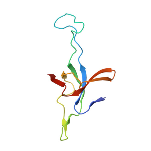
    Chaperonins are large barrel-shaped complexes that mediate ATP-dependent protein folding 1-3 . The bacterial chaperonin GroEL forms juxtaposed rings that bind unfolded protein and the lid-shaped cofactor GroES at their apertures. In vitro analyses of the chaperonin reaction have shown that substrate protein folds, unimpaired by aggregation, while transiently encapsulated in the GroEL central cavity by GroES 4-6 . To determine the functional stoichiometry of GroEL, GroES and client protein in situ, here we visualized chaperonin complexes in their natural cellular environment using cryo-electron tomography. We find that, under various growth conditions, around 55-70% of GroEL binds GroES asymmetrically on one ring, with the remainder populating symmetrical complexes. Bound substrate protein is detected on the free ring of the asymmetrical complex, defining the substrate acceptor state. In situ analysis of GroEL-GroES chambers, validated by high-resolution structures obtained in vitro, showed the presence of encapsulated substrate protein in a folded state before release into the cytosol. Based on a comprehensive quantification and conformational analysis of chaperonin complexes, we propose a GroEL-GroES reaction cycle that consists of linked asymmetrical and symmetrical subreactions mediating protein folding. Our findings illuminate the native conformational and functional chaperonin cycle directly within cells.


  • Organizational Affiliation
    • Department of Cellular Biochemistry, Max Planck Institute of Biochemistry, Martinsried, Germany.

Macromolecule Content 

  • Total Structure Weight: 955.25 kDa 
  • Atom Count: 64,400 
  • Modeled Residue Count: 8,666 
  • Deposited Residue Count: 9,016 
  • Unique protein chains: 2

Macromolecules

Find similar proteins by:|  3D Structure
Entity ID: 1
MoleculeChains  Sequence LengthOrganismDetailsImage
Chaperonin GroEL547Escherichia coli BL21(DE3)Mutation(s): 0 
Gene Names: groEL
Find similar proteins by:|  3D Structure
Entity ID: 2
MoleculeChains  Sequence LengthOrganismDetailsImage
Co-chaperonin GroES97Escherichia coli BL21(DE3)Mutation(s): 0 
Gene Names: groES

Small Molecules

Ligands 3 Unique
IDChains Name / Formula / InChI Key2D Diagram3D Interactions
ATP

Query on ATP


Download:Ideal Coordinates CCD File
AB [auth I]
CA [auth A]
DB [auth J]
FA [auth B]
GB [auth K]
AB [auth I],
CA [auth A],
DB [auth J],
FA [auth B],
GB [auth K],
IA [auth C],
JB [auth L],
LA [auth D],
MB [auth M],
OA [auth E],
PB [auth N],
RA [auth F],
UA [auth G],
XA [auth H]
ADENOSINE-5'-TRIPHOSPHATE
C10 H16 N5 O13 P3
ZKHQWZAMYRWXGA-KQYNXXCUSA-N
K

Query on K


Download:Ideal Coordinates CCD File
CB [auth I]
EA [auth A]
FB [auth J]
HA [auth B]
IB [auth K]
CB [auth I],
EA [auth A],
FB [auth J],
HA [auth B],
IB [auth K],
KA [auth C],
LB [auth L],
NA [auth D],
OB [auth M],
QA [auth E],
RB [auth N],
TA [auth F],
WA [auth G],
ZA [auth H]
POTASSIUM ION
K
NPYPAHLBTDXSSS-UHFFFAOYSA-N
MG

Query on MG


Download:Ideal Coordinates CCD File
BB [auth I]
DA [auth A]
EB [auth J]
GA [auth B]
HB [auth K]
BB [auth I],
DA [auth A],
EB [auth J],
GA [auth B],
HB [auth K],
JA [auth C],
KB [auth L],
MA [auth D],
NB [auth M],
PA [auth E],
QB [auth N],
SA [auth F],
VA [auth G],
YA [auth H]
MAGNESIUM ION
Mg
JLVVSXFLKOJNIY-UHFFFAOYSA-N

Experimental Data & Validation

Experimental Data

  • Method: ELECTRON MICROSCOPY
  • Resolution: 11.9 Å
  • Aggregation State: CELL 
  • Reconstruction Method: SUBTOMOGRAM AVERAGING 
EM Software:
TaskSoftware PackageVersion
RECONSTRUCTIONSTOPGAP

Structure Validation

View Full Validation Report


Entry History 

& Funding Information

Deposition Data


Funding OrganizationLocationGrant Number
Not funded--

Revision History  (Full details and data files)

  • Version 1.0: 2024-07-03
    Type: Initial release
  • Version 1.1: 2024-08-21
    Changes: Data collection, Database references
  • Version 1.2: 2024-09-04
    Changes: Data collection, Database references
  • Version 1.3: 2024-09-25
    Changes: Data collection, Database references