Probing a Major DNA Weakness: Resolving the Groove and Sequence Selectivity of the Diimine Complex Lambda-[Ru(phen) 2 phi] 2.
Prieto Otoya, T.D., McQuaid, K.T., Hennessy, J., Menounou, G., Gibney, A., Paterson, N.G., Cardin, D.J., Kellett, A., Cardin, C.J.(2024) Angew Chem Int Ed Engl 63: e202318863-e202318863
- PubMed: 38271265
- DOI: https://doi.org/10.1002/anie.202318863
- Primary Citation of Related Structures:
8OYR - PubMed Abstract:
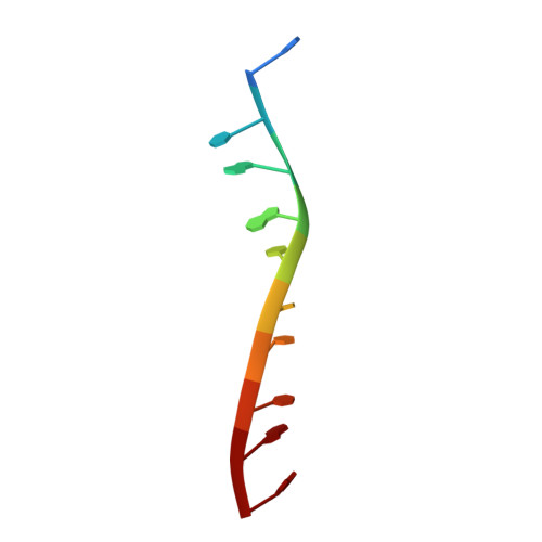
The grooves of DNA provide recognition sites for many nucleic acid binding proteins and anticancer drugs such as the covalently binding cisplatin. Here we report a crystal structure showing, for the first time, groove selectivity by an intercalating ruthenium complex. The complex Λ-[Ru(phen) 2 phi] 2+ , where phi=9,10-phenanthrenediimine, is bound to the DNA decamer duplex d(CCGGTACCGG) 2 . The structure shows that the metal complex is symmetrically bound in the major groove at the central TA/TA step, and asymmetrically bound in the minor groove at the adjacent GG/CC steps. A third type of binding links the strands, in which each terminal cytosine base stacks with one phen ligand. The overall binding stoichiometry is four Ru complexes per duplex. Complementary biophysical measurements confirm the binding preference for the Λ-enantiomer and show a high affinity for TA/TA steps and, more generally, TA-rich sequences. A striking enantiospecific elevation of melting temperatures is found for oligonucleotides which include the TATA box sequence.
- Department of Chemistry, University of Reading, Whiteknights, Reading, RG6 6AD, UK.
Organizational Affiliation:


