8OXD | pdb_00008oxd

Structure of the human LYVE-1 (lymphatic vessel endothelial receptor-1) hyaluronan binding domain bound with decasaccharide hyaluronan.


Experimental Data Snapshot

  • Method: X-RAY DIFFRACTION
  • Resolution: 1.32 Å
  • R-Value Free: 
    0.217 (Depositor), 0.216 (DCC) 
  • R-Value Work: 
    0.172 (Depositor), 0.172 (DCC) 
  • R-Value Observed: 
    0.175 (Depositor) 

Starting Model: experimental
View more details

wwPDB Validation   3D Report Full Report


Ligand Structure Quality Assessment 


This is version 1.1 of the entry. See complete history


Literature

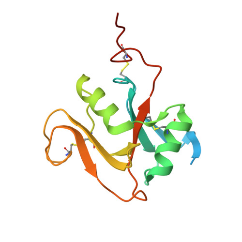
Structure of the murine LYVE-1 (lymphatic vessel endothelial receptor-1) hyaluronan binding domain in an unliganded state

Bano, F.Banerji, S.Ni, T.Green, D.E.DeAngelis, P.L.Paci, E.Lepsik, M.Jackson, D.J.Gilbert, R.J.C.

(2025) Nature Communications 


Macromolecules
Find similar proteins by:  (by identity cutoff)  |  3D Structure
Entity ID: 1
MoleculeChains Sequence LengthOrganismDetailsImage
Lymphatic vessel endothelial hyaluronic acid receptor 1146Homo sapiensMutation(s): 0 
Gene Names: LYVE1CRSBP1HARXLKD1UNQ230/PRO263
UniProt & NIH Common Fund Data Resources
Find proteins for Q9Y5Y7 (Homo sapiens)
Explore Q9Y5Y7 
Go to UniProtKB:  Q9Y5Y7
PHAROS:  Q9Y5Y7
GTEx:  ENSG00000133800 
Entity Groups  
Sequence Clusters30% Identity50% Identity70% Identity90% Identity95% Identity100% Identity
UniProt GroupQ9Y5Y7
Glycosylation
Glycosylation Sites: 3Go to GlyGen: Q9Y5Y7-1
Sequence Annotations
Expand
  • Reference Sequence
Oligosaccharides

Help

Entity ID: 2
MoleculeChains Length2D Diagram Glycosylation3D Interactions
2-acetamido-2-deoxy-beta-D-glucopyranose-(1-4)-beta-D-glucopyranuronic acid-(1-3)-2-acetamido-2-deoxy-beta-D-glucopyranose-(1-4)-beta-D-glucopyranuronic acid-(1-3)-2-acetamido-2-deoxy-beta-D-glucopyranose-(1-4)-beta-D-glucopyranuronic acidB [auth C]6N/A
Glycosylation Resources
GlyTouCan:  G97733LD
GlyCosmos:  G97733LD
GlyGen:  G97733LD
Experimental Data & Validation

Experimental Data

  • Method: X-RAY DIFFRACTION
  • Resolution: 1.32 Å
  • R-Value Free:  0.217 (Depositor), 0.216 (DCC) 
  • R-Value Work:  0.172 (Depositor), 0.172 (DCC) 
  • R-Value Observed: 0.175 (Depositor) 
Space Group: P 21 21 21
Unit Cell:
Length ( Å )Angle ( ˚ )
a = 39.59α = 90
b = 53.26β = 90
c = 53.74γ = 90
Software Package:
Software NamePurpose
PHENIXrefinement
xia2data reduction
xia2data scaling
PHENIXphasing
EM Software:
TaskSoftware PackageVersion
MODEL REFINEMENTPHENIX1.21rc1_4899

Structure Validation

View Full Validation Report


Ligand Structure Quality Assessment 


Entry History & Funding Information

Deposition Data


Funding OrganizationLocationGrant Number
Medical Research Council (MRC, United Kingdom)United Kingdom--

Revision History  (Full details and data files)

  • Version 1.0: 2024-11-13
    Type: Initial release
  • Version 1.1: 2025-03-26
    Changes: Database references, Structure summary