8OVS | pdb_00008ovs

Crystal structure of YeGT glycosyltransferase with bound UDP


Experimental Data Snapshot

  • Method: X-RAY DIFFRACTION
  • Resolution: 1.12 Å
  • R-Value Free: 
    0.169 (Depositor), 0.190 (DCC) 
  • R-Value Work: 
    0.153 (Depositor) 

Starting Model: experimental
View more details

wwPDB Validation 3D Report Full Report

Validation slider image for 8OVS

Ligand Structure Quality Assessment 


This is version 1.1 of the entry. See complete history

Literature

Tyrosine-modifying glycosylation by Yersinia effectors.

Schneider, S.Wirth, C.Jank, T.Hunte, C.Aktories, K.

(2024) J Biological Chem 300: 107331-107331

  • DOI: https://doi.org/10.1016/j.jbc.2024.107331
  • Primary Citation Related Structures: 
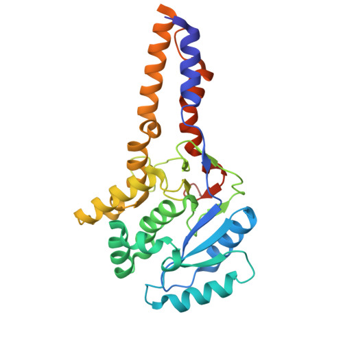
    8OVS, 8OVT

  • PubMed Abstract: 

    Mono-O-glycosylation of target proteins by bacterial toxins or effector proteins is a well-known mechanism by which bacteria interfere with essential functions of host cells. The respective glycosyltransferases are important virulence factors such as the Clostridioides difficile toxins A and B. Here, we describe two glycosyltransferases of Yersinia species that have a high sequence identity: YeGT from the zoonotic pathogen Yersinia enterocolitica and YkGT from the murine pathogen Yersinia kristensenii. We show that both modify Rho family proteins by attachment of N-acetylglucosamine (GlcNAc) at tyrosine residues (Tyr-34 in RhoA). Notably, the enzymes differed in their target protein specificity. While YeGT modified RhoA, B and C, YkGT possessed a broader substrate spectrum and glycosylated not only Rho but also Rac and Cdc42 subfamily proteins. Mutagenesis studies indicated that residue 177 is important for this broader target spectrum. We determined the crystal structure of YeGT shortened by 16 residues N-terminally (sYeGT) in the ligand-free state and bound to UDP, the product of substrate hydrolysis. The structure assigns sYeGT to the GT-A family. It shares high structural similarity to glycosyltransferase domains from toxins. We also demonstrated that the 16 most N-terminal residues of YeGT and YkGT are important for the mediated translocation into the host cell using the pore-forming protective antigen of anthrax toxin. Mediated introduction into Hela cells or ectopic expression of YeGT and YkGT caused morphological changes and redistribution of the actin cytoskeleton. The data suggest that YeGT and YkGT are likely bacterial effectors belonging to the family of tyrosine glycosylating bacterial glycosyltransferases.


  • Organizational Affiliation
    • Institute for Experimental and Clinical Pharmacology and Toxicology, Faculty of Medicine, University of Freiburg, D-79104 Freiburg, Germany.

Macromolecule Content 

  • Total Structure Weight: 71.73 kDa 
  • Atom Count: 5,565 
  • Modeled Residue Count: 547 
  • Deposited Residue Count: 598 
  • Unique protein chains: 1

Macromolecules

Find similar proteins by:|  3D Structure
Entity ID: 1
MoleculeChains  Sequence LengthOrganismDetailsImage
YeGT glycosyltransferaseA [auth AAA],
B [auth BBB]
299Yersinia enterocoliticaMutation(s): 0 

Small Molecules

Ligands 6 Unique
IDChains Name / Formula / InChI Key2D Diagram3D Interactions
UDP
(Subject of Investigation/LOI)

Query on UDP


Download:Ideal Coordinates CCD File
C [auth AAA],
Y [auth BBB]
URIDINE-5'-DIPHOSPHATE
C9 H14 N2 O12 P2
XCCTYIAWTASOJW-XVFCMESISA-N
CD

Query on CD


Download:Ideal Coordinates CCD File
GA [auth BBB]
HA [auth BBB]
IA [auth BBB]
J [auth AAA]
JA [auth BBB]
GA [auth BBB],
HA [auth BBB],
IA [auth BBB],
J [auth AAA],
JA [auth BBB],
K [auth AAA],
KA [auth BBB],
L [auth AAA],
LA [auth BBB],
M [auth AAA],
MA [auth BBB],
N [auth AAA],
O [auth AAA],
P [auth AAA],
Q [auth AAA],
R [auth AAA],
S [auth AAA]
CADMIUM ION
Cd
WLZRMCYVCSSEQC-UHFFFAOYSA-N
EDO

Query on EDO


Download:Ideal Coordinates CCD File
AA [auth BBB]
BA [auth BBB]
CA [auth BBB]
DA [auth BBB]
EA [auth BBB]
AA [auth BBB],
BA [auth BBB],
CA [auth BBB],
DA [auth BBB],
EA [auth BBB],
F [auth AAA],
FA [auth BBB],
G [auth AAA],
I [auth AAA],
Z [auth BBB]
1,2-ETHANEDIOL
C2 H6 O2
LYCAIKOWRPUZTN-UHFFFAOYSA-N
ACT

Query on ACT


Download:Ideal Coordinates CCD File
D [auth AAA],
E [auth AAA],
H [auth AAA],
W [auth BBB],
X [auth BBB]
ACETATE ION
C2 H3 O2
QTBSBXVTEAMEQO-UHFFFAOYSA-M
MN
(Subject of Investigation/LOI)

Query on MN


Download:Ideal Coordinates CCD File
NA [auth BBB],
T [auth AAA]
MANGANESE (II) ION
Mn
WAEMQWOKJMHJLA-UHFFFAOYSA-N
NA

Query on NA


Download:Ideal Coordinates CCD File
OA [auth BBB],
U [auth AAA],
V [auth AAA]
SODIUM ION
Na
FKNQFGJONOIPTF-UHFFFAOYSA-N

Experimental Data & Validation

Experimental Data

  • Method: X-RAY DIFFRACTION
  • Resolution: 1.12 Å
  • R-Value Free:  0.169 (Depositor), 0.190 (DCC) 
  • R-Value Work:  0.153 (Depositor) 
Space Group: P 1 21 1
Unit Cell:
Length ( Å )Angle ( ˚ )
a = 47.13α = 90
b = 114.546β = 89.987
c = 69.376γ = 90
Software Package:
Software NamePurpose
REFMACrefinement
MxCuBEdata collection
autoPROCdata processing
PHASERphasing
STARANISOdata scaling
Cootmodel building
autoPROCdata reduction

Structure Validation

View Full Validation Report


Ligand Structure Quality Assessment 


Entry History 

& Funding Information

Deposition Data


Funding OrganizationLocationGrant Number
German Research Foundation (DFG)GermanyCRC 746
German Federal Ministry for Education and ResearchGermanyEXC-2189

Revision History  (Full details and data files)

  • Version 1.0: 2024-05-22
    Type: Initial release
  • Version 1.1: 2024-06-05
    Changes: Database references