


The crystal structure of PET44, a PETase enzyme from Alkalilimnicola ehrlichii
Costanzi, E., Applegate, V., Smits, S.H.J.To be published.
Experimental Data Snapshot
Starting Model: in silico
View more details
wwPDB Validation 3D Report Full Report

Entity ID: 1 | |||||
|---|---|---|---|---|---|
| Molecule | Chains | Sequence Length | Organism | Details | Image |
| Dienelactone hydrolase domain-containing protein | 320 | Alkalilimnicola ehrlichii | Mutation(s): 0 Gene Names: CAL65_11960 | 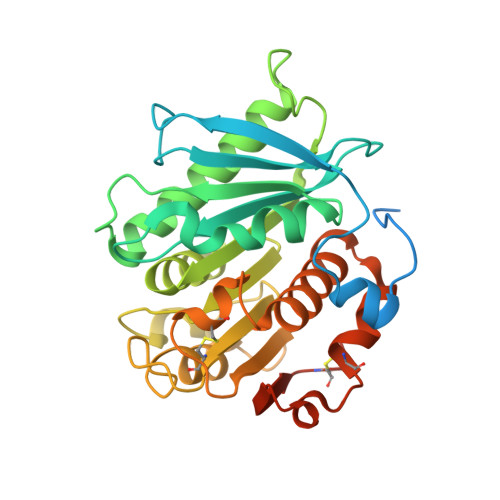 | |
UniProt | |||||
Find proteins for A0A3E0WVY1 (Alkalilimnicola ehrlichii) Explore A0A3E0WVY1 Go to UniProtKB: A0A3E0WVY1 | |||||
Entity Groups | |||||
| Sequence Clusters | 30% Identity50% Identity70% Identity90% Identity95% Identity100% Identity | ||||
| UniProt Group | A0A3E0WVY1 | ||||
Sequence AnnotationsExpand | |||||
Reference Sequence | |||||
| Ligands 3 Unique | |||||
|---|---|---|---|---|---|
| ID | Chains | Name / Formula / InChI Key | 2D Diagram | 3D Interactions | |
| EDO Download:Ideal Coordinates CCD File | AA [auth B] BA [auth B] CA [auth B] D [auth A] DA [auth B] | 1,2-ETHANEDIOL C2 H6 O2 LYCAIKOWRPUZTN-UHFFFAOYSA-N |  | ||
| CL Download:Ideal Coordinates CCD File | GA [auth B], T [auth A], U [auth A] | CHLORIDE ION Cl VEXZGXHMUGYJMC-UHFFFAOYSA-M |  | ||
| NA Download:Ideal Coordinates CCD File | C [auth A], V [auth B], W [auth B] | SODIUM ION Na FKNQFGJONOIPTF-UHFFFAOYSA-N |  | ||
| Length ( Å ) | Angle ( ˚ ) |
|---|---|
| a = 141.133 | α = 90 |
| b = 45.66 | β = 97.83 |
| c = 84.187 | γ = 90 |
| Software Name | Purpose |
|---|---|
| PHENIX | refinement |
| PHENIX | refinement |
| XDS | data reduction |
| Aimless | data scaling |
| PHASER | phasing |
| Funding Organization | Location | Grant Number |
|---|---|---|
| German Research Foundation (DFG) | Germany | 417919780 |
| German Federal Ministry for Education and Research | Germany | 31B0837A |


