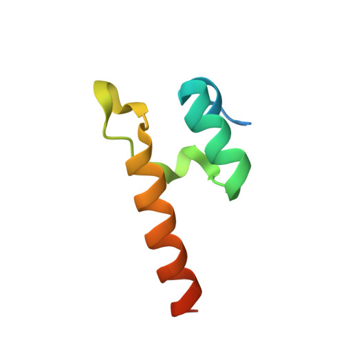
8OQJ

Peripheral subunit binding domain of the E. coli Dihydrolipoamide Acetyltransferase (E2) of the pyruvate dehydrogenase complex


Experimental Data Snapshot

  • Method: X-RAY DIFFRACTION
  • Resolution: 1.64 Å
  • R-Value Free: 0.245 
  • R-Value Work: 0.215 
  • R-Value Observed: 0.217 

wwPDB Validation   3D Report Full Report


This is version 1.1 of the entry. See complete history


Literature

Dimerization of a 5-kDa domain defines the architecture of the 5-MDa gammaproteobacterial pyruvate dehydrogenase complex.

Meinhold, S.Zdanowicz, R.Giese, C.Glockshuber, R.

(2024) Sci Adv 10: eadj6358-eadj6358

  • DOI: https://doi.org/10.1126/sciadv.adj6358
  • Primary Citation of Related Structures:  
    8OQJ, 8ORB, 8OSY

  • PubMed Abstract: 

    The Escherichia coli pyruvate dehydrogenase complex (PDHc) is a ~5 MDa assembly of the catalytic subunits pyruvate dehydrogenase (E1), dihydrolipoamide acetyltransferase (E2), and dihydrolipoamide dehydrogenase (E3). The PDHc core is a cubic complex of eight E2 homotrimers. Homodimers of the peripheral subunits E1 and E3 associate with the core by binding to the peripheral subunit binding domain (PSBD) of E2. Previous reports indicated that 12 E1 dimers and 6 E3 dimers bind to the 24-meric E2 core. Using an assembly arrested E2 homotrimer (E2 3 ), we show that two of the three PSBDs in the E2 3 dimerize, that each PSBD dimer cooperatively binds two E1 dimers, and that E3 dimers only bind to the unpaired PSBD in E2 3 . This mechanism is preserved in wild-type PDHc, with an E1 dimer:E2 monomer:E3 dimer stoichiometry of 16:24:8. The conserved PSBD dimer interface indicates that PSBD dimerization is the previously unrecognized architectural determinant of gammaproteobacterial PDHc megacomplexes.


  • Organizational Affiliation

    ETH Zürich, Institute of Molecular Biology and Biophysics, Otto-Stern-Weg 5, 8093 Zürich, Switzerland.


Macromolecules
Find similar proteins by:  (by identity cutoff)  |  3D Structure
Entity ID: 1
MoleculeChains Sequence LengthOrganismDetailsImage
Dihydrolipoyllysine-residue acetyltransferase component of pyruvate dehydrogenase complex
A, B
67Escherichia coliMutation(s): 0 
Gene Names: aceFb0115JW0111
EC: 2.3.1.12
UniProt
Find proteins for P06959 (Escherichia coli (strain K12))
Explore P06959 
Go to UniProtKB:  P06959
Entity Groups  
Sequence Clusters30% Identity50% Identity70% Identity90% Identity95% Identity100% Identity
UniProt GroupP06959
Sequence Annotations
Expand
  • Reference Sequence
Small Molecules
Ligands 1 Unique
IDChains Name / Formula / InChI Key2D Diagram3D Interactions
ZN
Query on ZN

Download Ideal Coordinates CCD File 
C [auth B]ZINC ION
Zn
PTFCDOFLOPIGGS-UHFFFAOYSA-N
Experimental Data & Validation

Experimental Data

  • Method: X-RAY DIFFRACTION
  • Resolution: 1.64 Å
  • R-Value Free: 0.245 
  • R-Value Work: 0.215 
  • R-Value Observed: 0.217 
  • Space Group: P 1
Unit Cell:
Length ( Å )Angle ( ˚ )
a = 33.576α = 100.39
b = 37.831β = 114.386
c = 37.834γ = 101.179
Software Package:
Software NamePurpose
PHENIXrefinement
XDSdata reduction
XDSdata scaling
PHASERphasing

Structure Validation

View Full Validation Report


Entry History & Funding Information

Deposition Data


Funding OrganizationLocationGrant Number
Swiss National Science FoundationSwitzerland--

Revision History  (Full details and data files)

  • Version 1.0: 2024-02-07
    Type: Initial release
  • Version 1.1: 2024-02-21
    Changes: Database references