8OOY

Pol I bound to extended and displaced DNA section - open conformation


Experimental Data Snapshot

  • Method: ELECTRON MICROSCOPY
  • Resolution: 4.00 Å
  • Aggregation State: PARTICLE 
  • Reconstruction Method: SINGLE PARTICLE 

wwPDB Validation   3D Report Full Report


This is version 1.2 of the entry. See complete history


Literature

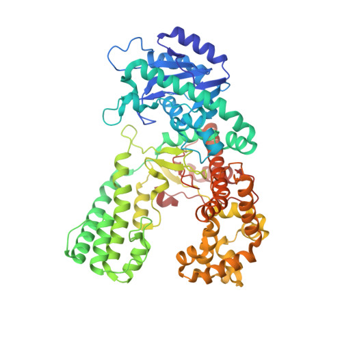
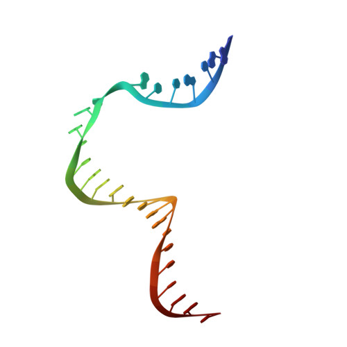
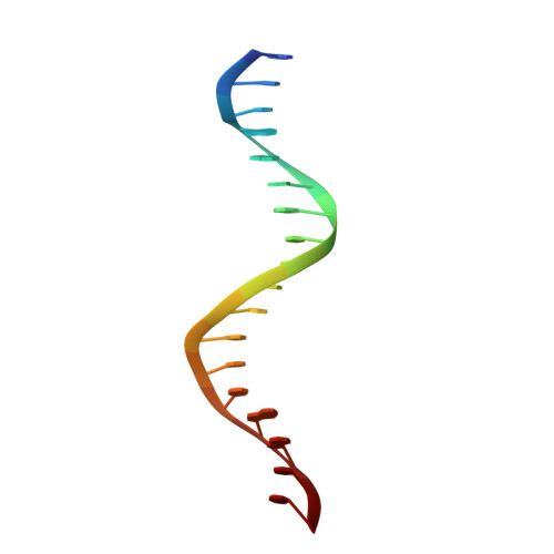
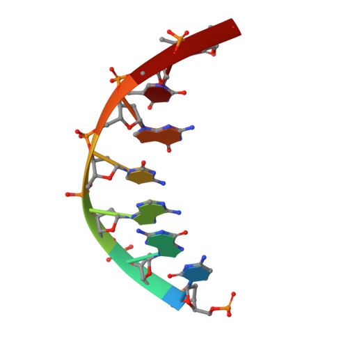
A four-point molecular handover during Okazaki maturation.

Botto, M.M.Borsellini, A.Lamers, M.H.

(2023) Nat Struct Mol Biol 30: 1505-1515

  • DOI: https://doi.org/10.1038/s41594-023-01071-y
  • Primary Citation of Related Structures:  
    8OO6, 8OOY

  • PubMed Abstract: 

    DNA replication introduces thousands of RNA primers into the lagging strand that need to be removed for replication to be completed. In Escherichia coli when the replicative DNA polymerase Pol IIIα terminates at a previously synthesized RNA primer, DNA Pol I takes over and continues DNA synthesis while displacing the downstream RNA primer. The displaced primer is subsequently excised by an endonuclease, followed by the sealing of the nick by a DNA ligase. Yet how the sequential actions of Pol IIIα, Pol I polymerase, Pol I endonuclease and DNA ligase are coordinated is poorly defined. Here we show that each enzymatic activity prepares the DNA substrate for the next activity, creating an efficient four-point molecular handover. The cryogenic-electron microscopy structure of Pol I bound to a DNA substrate with both an upstream and downstream primer reveals how it displaces the primer in a manner analogous to the monomeric helicases. Moreover, we find that in addition to its flap-directed nuclease activity, the endonuclease domain of Pol I also specifically cuts at the RNA-DNA junction, thus marking the end of the RNA primer and creating a 5' end that is a suitable substrate for the ligase activity of LigA once all RNA has been removed.


  • Organizational Affiliation

    Department of Cell and Chemical Biology, Leiden University Medical Center (LUMC), Leiden, the Netherlands.


Macromolecules

Find similar proteins by:  (by identity cutoff)  |  3D Structure
Entity ID: 1
MoleculeChains Sequence LengthOrganismDetailsImage
DNA polymerase I604Escherichia coli 'BL21-Gold(DE3)pLysS AGMutation(s): 0 
Gene Names: polAresAb3863JW3835
EC: 2.7.7.7
UniProt
Find proteins for P00582 (Escherichia coli (strain K12))
Explore P00582 
Go to UniProtKB:  P00582
Entity Groups  
Sequence Clusters30% Identity50% Identity70% Identity90% Identity95% Identity100% Identity
UniProt GroupP00582
Sequence Annotations
Expand
  • Reference Sequence
Find similar nucleic acids by: 3D Structure
Entity ID: 2
MoleculeChains LengthOrganismImage
Template DNAB [auth T]26synthetic construct
Sequence Annotations
Expand
  • Reference Sequence

Find similar nucleic acids by:  Sequence   |   3D Structure  

Entity ID: 3
MoleculeChains LengthOrganismImage
Extending PrimerC [auth P]18synthetic construct
Sequence Annotations
Expand
  • Reference Sequence

Find similar nucleic acids by:  Sequence   |   3D Structure  

Entity ID: 4
MoleculeChains LengthOrganismImage
Displacing Primer7synthetic construct
Sequence Annotations
Expand
  • Reference Sequence
Small Molecules
Ligands 1 Unique
IDChains Name / Formula / InChI Key2D Diagram3D Interactions
MG
Query on MG

Download Ideal Coordinates CCD File 
E [auth A]MAGNESIUM ION
Mg
JLVVSXFLKOJNIY-UHFFFAOYSA-N
Experimental Data & Validation

Experimental Data

  • Method: ELECTRON MICROSCOPY
  • Resolution: 4.00 Å
  • Aggregation State: PARTICLE 
  • Reconstruction Method: SINGLE PARTICLE 

Structure Validation

View Full Validation Report


Entry History & Funding Information

Deposition Data


Funding OrganizationLocationGrant Number
Other government--

Revision History  (Full details and data files)

  • Version 1.0: 2023-08-09
    Type: Initial release
  • Version 1.1: 2023-09-06
    Changes: Data collection, Database references
  • Version 1.2: 2023-11-01
    Changes: Database references