Cisplatin binding to angiogenin protein: new molecular pathways and targets for the drug's anticancer activity.
Ferraro, G., Sanfilippo, V., Chiaverini, L., Satriano, C., Marzo, T., Merlino, A., La Mendola, D.(2023) Dalton Trans 52: 9058-9067
- PubMed: 37337706 Search on PubMed
- DOI: https://doi.org/10.1039/d3dt01517c
- Primary Citation Related Structures:
8OO3, 8OO4 - PubMed Abstract:
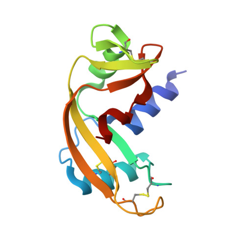
Cisplatin (CisPt), a platinum-based chemotherapeutic widely used in the treatment of various cancers, has multiple mechanisms of action, including nuclear DNA (nDNA) and mitochondrial DNA (mDNA) damage and cytoskeletal perturbations affecting, in turn, the membrane transporter activity. CisPt binding to proteins and enzymes may modulate its biochemical mechanism of action and is associated with cancer cell resistance to the drug. In this work, we investigate the interaction between cisplatin and angiogenin (Ang), a protein strongly expressed in many types of cancer and a potent angiogenic factor. The adduct formed upon reaction of CisPt with Ang (Ang@CisPt) was characterized by X-ray crystallography to evidence the exact platination site and by UV-visible (UV-vis) absorption and circular dichroism (CD) spectroscopies to shed light on any possible change in the protein conformation. Furthermore, high-resolution electrospray ionization (ESI) mass spectrometry was utilized to evaluate the Ang : CisPt stoichiometry of the Ang@CisPt adduct. The effect of the Ang@CisPt adduct on a prostate cancer cell line (PC-3) was tested by colorimetric assays in terms of cell viability, at both levels of nuclear and mitochondrial damage, and reactive oxygen species (ROS) production. Cellular imaging by laser scanning confocal microscopy (LSM) was utilized to scrutinize the cytoskeleton actin reorganization and the lysosome and mitochondria organelle perturbation. These studies highlight the possibility of new molecular pathways and targets for CisPt activity.
- Department of Chemical Sciences, University of Naples Federico II, via Cintia 21, I-80126 Napoli, Italy. antonello.merlino@unina.it.
Organizational Affiliation: