Molecular mechanism of IKK catalytic dimer docking to NF-kappa B substrates.
Li, C., Moro, S., Shostak, K., O'Reilly, F.J., Donzeau, M., Graziadei, A., McEwen, A.G., Desplancq, D., Poussin-Courmontagne, P., Bachelart, T., Fiskin, M., Berrodier, N., Pichard, S., Brillet, K., Orfanoudakis, G., Poterszman, A., Torbeev, V., Rappsilber, J., Davey, N.E., Chariot, A., Zanier, K.(2024) Nat Commun 15: 7692-7692
- PubMed: 39227404 Search on PubMedSearch on PubMed Central
- DOI: https://doi.org/10.1038/s41467-024-52076-0
- Primary Citation Related Structures:
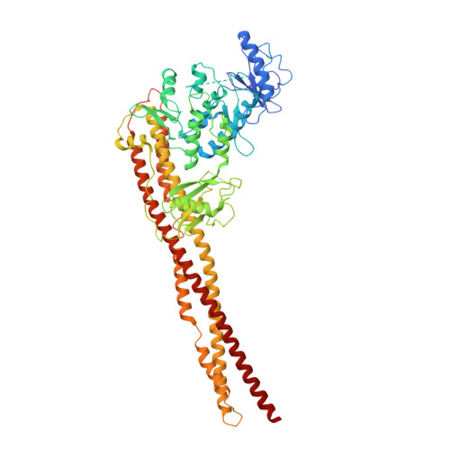
8OMV - PubMed Abstract:
The inhibitor of κB (IκB) kinase (IKK) is a central regulator of NF-κB signaling. All IKK complexes contain hetero- or homodimers of the catalytic IKKβ and/or IKKα subunits. Here, we identify a YDDΦxΦ motif, which is conserved in substrates of canonical (IκBα, IκBβ) and alternative (p100) NF-κB pathways, and which mediates docking to catalytic IKK dimers. We demonstrate a quantitative correlation between docking affinity and IKK activity related to IκBα phosphorylation/degradation. Furthermore, we show that phosphorylation of the motif's conserved tyrosine, an event previously reported to promote IκBα accumulation and inhibition of NF-κB gene expression, suppresses the docking interaction. Results from integrated structural analyzes indicate that the motif binds to a groove at the IKK dimer interface. Consistently, suppression of IKK dimerization also abolishes IκBα substrate binding. Finally, we show that an optimized bivalent motif peptide inhibits NF-κB signaling. This work unveils a function for IKKα/β dimerization in substrate motif recognition.
- Biotechnology and Cell Signaling (CNRS/Université de Strasbourg, UMR7242), Ecole Superieure de Biotechnologie de Strasbourg, Boulevard Sébastien Brant, 67400, Illkirch, France.
Organizational Affiliation: