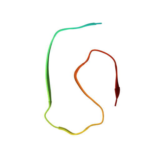
Cryo-EM of A beta fibrils from mouse models find tg-APP ArcSwe fibrils resemble those found in patients with sporadic Alzheimer's disease.
Zielinski, M., Peralta Reyes, F.S., Gremer, L., Schemmert, S., Frieg, B., Schafer, L.U., Willuweit, A., Donner, L., Elvers, M., Nilsson, L.N.G., Syvanen, S., Sehlin, D., Ingelsson, M., Willbold, D., Schroder, G.F.(2023) Nat Neurosci 26: 2073-2080
- PubMed: 37973869 Search on PubMedSearch on PubMed Central
- DOI: https://doi.org/10.1038/s41593-023-01484-4
- Primary Citation Related Structures:
8OL2, 8OL3, 8OL5, 8OL6, 8OL7, 8OLG, 8OLN, 8OLO, 8OLQ - PubMed Abstract:
The use of transgenic mice displaying amyloid-β (Aβ) brain pathology has been essential for the preclinical assessment of new treatment strategies for Alzheimer's disease. However, the properties of Aβ in such mice have not been systematically compared to Aβ in the brains of patients with Alzheimer's disease. Here, we determined the structures of nine ex vivo Aβ fibrils from six different mouse models by cryogenic-electron microscopy. We found novel Aβ fibril structures in the APP/PS1, ARTE10 and tg-SwDI models, whereas the human type II filament fold was found in the ARTE10, tg-APP Swe and APP23 models. The tg-APP ArcSwe mice showed an Aβ fibril whose structure resembles the human type I filament found in patients with sporadic Alzheimer's disease. A detailed assessment of the Aβ fibril structure is key to the selection of adequate mouse models for the preclinical development of novel plaque-targeting therapeutics and positron emission tomography imaging tracers in Alzheimer's disease.
- Institute of Biological Information Processing, Structural Biochemistry (IBI-7), Forschungszentrum Jülich, Jülich, Germany.
Organizational Affiliation: