


Structure of Mycobacterium tuberculosis 1-Deoxy-D-Xylulose 5-Phosphate Synthase in Complex with Butylacetylphosphonate
Gawriljuk, V.O., Oerlemans, R., Gierse, R.M., Jotwani, R., Hirsch, A.K.H., Groves, M.R.(2023) Crystals (Basel) 13
Experimental Data Snapshot
Starting Model: experimental
View more details
(2023) Crystals (Basel) 13
Entity ID: 1 | |||||
|---|---|---|---|---|---|
| Molecule | Chains | Sequence Length | Organism | Details | Image |
| 1-deoxy-D-xylulose-5-phosphate synthase | 604 | Mycobacterium tuberculosis H37Rv | Mutation(s): 0 Gene Names: dxs, Rv2682c, MTCY05A6.03c EC: 2.2.1.7 | 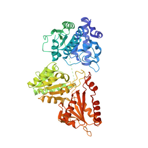 | |
UniProt | |||||
Entity Groups | |||||
| Sequence Clusters | 30% Identity50% Identity70% Identity90% Identity95% Identity100% Identity | ||||
| UniProt Group | P9WNS3 | ||||
Sequence AnnotationsExpand | |||||
Reference Sequence | |||||
| Ligands 5 Unique | |||||
|---|---|---|---|---|---|
| ID | Chains | Name / Formula / InChI Key | 2D Diagram | 3D Interactions | |
| VMI (Subject of Investigation/LOI) Download:Ideal Coordinates CCD File | C [auth A], M [auth B] | [(S)-1-[3-[(4-azanyl-2-methyl-pyrimidin-5-yl)methyl]-4-methyl-5-[2-[oxidanyl(phosphonooxy)phosphoryl]oxyethyl]-1,3-thiazol-3-ium-2-yl]-1-oxidanyl-ethyl]-butoxy-phosphinic acid C18 H32 N4 O11 P3 S KQPODQFTFSEDTF-SFHVURJKSA-O |  | ||
| PEG Download:Ideal Coordinates CCD File | Q [auth B] | DI(HYDROXYETHYL)ETHER C4 H10 O3 MTHSVFCYNBDYFN-UHFFFAOYSA-N |  | ||
| GOL Download:Ideal Coordinates CCD File | D [auth A] | GLYCEROL C3 H8 O3 PEDCQBHIVMGVHV-UHFFFAOYSA-N |  | ||
| EDO Download:Ideal Coordinates CCD File | E [auth A] F [auth A] G [auth A] H [auth A] I [auth A] | 1,2-ETHANEDIOL C2 H6 O2 LYCAIKOWRPUZTN-UHFFFAOYSA-N |  | ||
| MG Download:Ideal Coordinates CCD File | L [auth A], R [auth B] | MAGNESIUM ION Mg JLVVSXFLKOJNIY-UHFFFAOYSA-N |  | ||
| Length ( Å ) | Angle ( ˚ ) |
|---|---|
| a = 63.462 | α = 90 |
| b = 127.775 | β = 106.883 |
| c = 79.583 | γ = 90 |
| Software Name | Purpose |
|---|---|
| PHENIX | refinement |
| XDS | data reduction |
| Aimless | data scaling |
| PHASER | phasing |
| Funding Organization | Location | Grant Number |
|---|---|---|
| H2020 Marie Curie Actions of the European Commission | European Union | 860816 |


