8OG0 | pdb_00008og0

Crystal structure of MJF14-6-4-2 Fab fragment in complex with epitope peptide


Experimental Data Snapshot

  • Method: X-RAY DIFFRACTION
  • Resolution: 1.71 Å
  • R-Value Free: 
    0.231 (Depositor), 0.236 (DCC) 
  • R-Value Work: 
    0.195 (Depositor), 0.204 (DCC) 
  • R-Value Observed: 
    0.197 (Depositor) 

Starting Model: experimental
View more details

wwPDB Validation 3D Report Full Report

Validation slider image for 8OG0

This is version 1.1 of the entry. See complete history

Literature

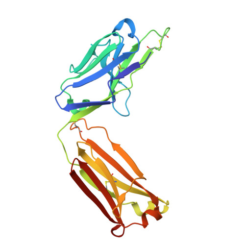
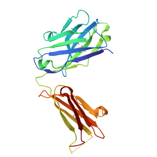
Structural basis of epitope recognition by anti-alpha-synuclein antibodies MJFR14-6-4-2.

Lieknina, I.Reimer, L.Pantelejevs, T.Lends, A.Jaudzems, K.El-Turabi, A.Gram, H.Hammi, A.Jensen, P.H.Tars, K.

(2024) NPJ Parkinsons Dis 10: 206-206

  • DOI: https://doi.org/10.1038/s41531-024-00822-y
  • Primary Citation Related Structures: 
    8OG0

  • PubMed Abstract: 

    Alpha-synuclein (α-syn) inclusions in the brain are hallmarks of so-called Lewy body diseases. Lewy bodies contain mainly aggregated α-syn together with some other proteins. Monomeric α-syn lacks a well-defined three-dimensional structure, but it can aggregate into oligomeric and fibrillar amyloid species, which can be detected using specific antibodies. Here we investigate the aggregate specificity of monoclonal MJFR14-6-4-2 antibodies. We conclude that partial masking of epitope in unstructured monomer in combination with a high local concentration of epitopes is the main reason for MJFR14-6-4-2 selectivity towards aggregates. Based on the structural insight, we produced mutant α-syn that when fibrillated is unable to bind MJFR14-6-4-2. Using these fibrils as a tool for seeding cellular α-syn aggregation, provides superior signal/noise ratio for detection of cellular α-syn aggregates by MJFR14-6-4-2. Our data provide a molecular level understanding of specific recognition of toxic amyloid oligomers, which is critical for the development of inhibitors against synucleinopathies.


  • Organizational Affiliation
    • Latvian Biomedical Research and Study Centre, Ratsupites 1, k-1, LV-1067, Riga, Latvia.

Macromolecule Content 

  • Total Structure Weight: 46.86 kDa 
  • Atom Count: 3,447 
  • Modeled Residue Count: 432 
  • Deposited Residue Count: 443 
  • Unique protein chains: 3

Macromolecules

Find similar proteins by:|  3D Structure
Entity ID: 1
MoleculeChains  Sequence LengthOrganismDetailsImage
Fab fragment light chainA [auth L]216Oryctolagus cuniculusMutation(s): 0 
Find similar proteins by:|  3D Structure
Entity ID: 2
MoleculeChains  Sequence LengthOrganismDetailsImage
Fab fragment heavy chainB [auth H]222Oryctolagus cuniculusMutation(s): 0 
Find similar proteins by:  Sequence   |   3D Structure  
Entity ID: 3
MoleculeChains  Sequence LengthOrganismDetailsImage
Alpha-synucleinC [auth P]5Homo sapiensMutation(s): 0 
UniProt & NIH Common Fund Data Resources
Find proteins for P37840 (Homo sapiens)
Explore P37840 
Go to UniProtKB:  P37840
PHAROS:  P37840
GTEx:  ENSG00000145335 
Entity Groups
UniProt GroupP37840
Sequence Annotations
Expand
Reference Sequence

Experimental Data & Validation

Experimental Data

  • Method: X-RAY DIFFRACTION
  • Resolution: 1.71 Å
  • R-Value Free:  0.231 (Depositor), 0.236 (DCC) 
  • R-Value Work:  0.195 (Depositor), 0.204 (DCC) 
  • R-Value Observed: 0.197 (Depositor) 
Space Group: P 21 21 21
Unit Cell:
Length ( Å )Angle ( ˚ )
a = 37.484α = 90
b = 66.7β = 90
c = 167.167γ = 90
Software Package:
Software NamePurpose
REFMACrefinement
autoPROCdata reduction
autoPROCdata scaling
PHASERphasing

Structure Validation

View Full Validation Report


Entry History 

& Funding Information

Deposition Data


Funding OrganizationLocationGrant Number
European Union (EU)European UnionOligoFIT

Revision History  (Full details and data files)

  • Version 1.0: 2024-03-27
    Type: Initial release
  • Version 1.1: 2024-11-06
    Changes: Database references, Structure summary