8K9O | pdb_00008k9o

Crystal structure of Cyanobacteriochrome RcaE GAF domain in Pg state


Experimental Data Snapshot

  • Method: X-RAY DIFFRACTION
  • Resolution: 1.75 Å
  • R-Value Free: 
    0.231 (Depositor), 0.242 (DCC) 
  • R-Value Work: 
    0.188 (Depositor), 0.198 (DCC) 

Starting Model: experimental
View more details

wwPDB Validation 3D Report Full Report

Validation slider image for 8K9O

Ligand Structure Quality Assessment 


This is version 1.2 of the entry. See complete history

Literature

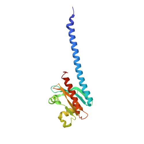
Green/red light-sensing mechanism in the chromatic acclimation photosensor.

Nagae, T.Fujita, Y.Tsuchida, T.Kamo, T.Seto, R.Hamada, M.Aoyama, H.Sato-Tomita, A.Fujisawa, T.Eki, T.Miyanoiri, Y.Ito, Y.Soeta, T.Ukaji, Y.Unno, M.Mishima, M.Hirose, Y.

(2024) Sci Adv 10: eadn8386-eadn8386

  • DOI: https://doi.org/10.1126/sciadv.adn8386
  • Primary Citation Related Structures: 
    8K9O

  • PubMed Abstract: 

    Certain cyanobacteria alter their photosynthetic light absorption between green and red, a phenomenon called complementary chromatic acclimation. The acclimation is regulated by a cyanobacteriochrome-class photosensor that reversibly photoconverts between green-absorbing (Pg) and red-absorbing (Pr) states. Here, we elucidated the structural basis of the green/red photocycle. In the Pg state, the bilin chromophore adopted the extended C15- Z , anti structure within a hydrophobic pocket. Upon photoconversion to the Pr state, the bilin is isomerized to the cyclic C15- E , syn structure, forming a water channel in the pocket. The solvation/desolvation of the bilin causes changes in the protonation state and the stability of π-conjugation at the B ring, leading to a large absorption shift. These results advance our understanding of the enormous spectral diversity of the phytochrome superfamily.


  • Organizational Affiliation
    • Department of Molecular Biophysics, School of Pharmacy, Tokyo University of Pharmacy and Life Sciences, Hachioji, Tokyo 192-0392, Japan.

Macromolecule Content 

  • Total Structure Weight: 22.63 kDa 
  • Atom Count: 1,567 
  • Modeled Residue Count: 179 
  • Deposited Residue Count: 194 
  • Unique protein chains: 1

Macromolecules

Find similar proteins by:|  3D Structure
Entity ID: 1
MoleculeChains  Sequence LengthOrganismDetailsImage
histidine kinase194Microchaete diplosiphonMutation(s): 0 
Gene Names: rcaE
EC: 2.7.13.3
UniProt
Find proteins for Q47897 (Microchaete diplosiphon)
Explore Q47897 
Go to UniProtKB:  Q47897
Entity Groups
Sequence Clusters30% Identity50% Identity70% Identity90% Identity95% Identity100% Identity
UniProt GroupQ47897
Sequence Annotations
Expand
Reference Sequence

Small Molecules

Ligands 1 Unique
IDChains Name / Formula / InChI Key2D Diagram3D Interactions
CYC
(Subject of Investigation/LOI)

Query on CYC


Download:Ideal Coordinates CCD File
B [auth A]PHYCOCYANOBILIN
C33 H40 N4 O6
VXTXPYZGDQPMHK-GMXXPEQVSA-N

Experimental Data & Validation

Experimental Data

  • Method: X-RAY DIFFRACTION
  • Resolution: 1.75 Å
  • R-Value Free:  0.231 (Depositor), 0.242 (DCC) 
  • R-Value Work:  0.188 (Depositor), 0.198 (DCC) 
Space Group: P 43 21 2
Unit Cell:
Length ( Å )Angle ( ˚ )
a = 64.273α = 90
b = 64.273β = 90
c = 84.116γ = 90
Software Package:
Software NamePurpose
REFMACrefinement
XDSdata reduction
Aimlessdata scaling
MOLREPphasing

Structure Validation

View Full Validation Report


Ligand Structure Quality Assessment 


Entry History 

& Funding Information

Deposition Data


Funding OrganizationLocationGrant Number
Japan Society for the Promotion of Science (JSPS)Japan22H02562
Japan Society for the Promotion of Science (JSPS)Japan19K06707
Other privateJapan--

Revision History  (Full details and data files)

  • Version 1.0: 2024-05-15
    Type: Initial release
  • Version 1.1: 2024-06-26
    Changes: Database references
  • Version 1.2: 2024-11-13
    Changes: Structure summary