


Molecular Basis for the P450-Catalyzed sp3 C-N Glycosidic Bond Formation in Staurosporine Biosynthesis.
Xiao, F., Dong, S., Feng, Y., Li, W.(2024) ACS Catal 14: 14274-14284
Experimental Data Snapshot
Entity ID: 1 | |||||
|---|---|---|---|---|---|
| Molecule | Chains | Sequence Length | Organism | Details | Image |
| Cytochrome P450 | 440 | Streptomyces sanyensis | Mutation(s): 0 Gene Names: spcN | 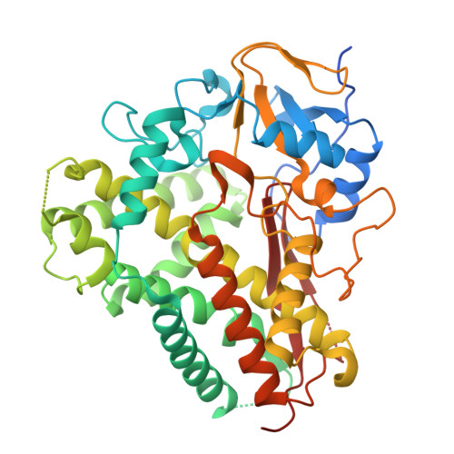 | |
UniProt | |||||
Entity Groups | |||||
| Sequence Clusters | 30% Identity50% Identity70% Identity90% Identity95% Identity100% Identity | ||||
| UniProt Group | W5R4Y8 | ||||
Sequence AnnotationsExpand | |||||
Reference Sequence | |||||
| Ligands 2 Unique | |||||
|---|---|---|---|---|---|
| ID | Chains | Name / Formula / InChI Key | 2D Diagram | 3D Interactions | |
| HEM (Subject of Investigation/LOI) Download:Ideal Coordinates CCD File | D [auth A], G [auth B], K [auth C] | PROTOPORPHYRIN IX CONTAINING FE C34 H32 Fe N4 O4 KABFMIBPWCXCRK-RGGAHWMASA-L |  | ||
| VI4 (Subject of Investigation/LOI) Download:Ideal Coordinates CCD File | E [auth A] F [auth A] H [auth B] I [auth B] J [auth B] | 13-(3-Amino-2,3,6-trideoxy-alpha-L-ribo-hexopyranosyl)-6,7,12,13-tetrahydro-5H-indolo[2,3-a]pyrrolo[3,4-c]carbazol-5-one C26 H24 N4 O3 QNQXRLNOOGRQEB-FUJFFIKUSA-N |  | ||
| Length ( Å ) | Angle ( ˚ ) |
|---|---|
| a = 206.96 | α = 90 |
| b = 121.79 | β = 91.45 |
| c = 64.21 | γ = 90 |
| Software Name | Purpose |
|---|---|
| PHENIX | refinement |
| xia2 | data scaling |
| xia2 | data reduction |
| PHENIX | phasing |
| Funding Organization | Location | Grant Number |
|---|---|---|
| National Natural Science Foundation of China (NSFC) | China | 32100051,32070054,U22A20582 |


