8JYC

Crystal Structure of Intracellular B30.2 Domain of BTN3A1 and BTN2A1 in Complex with DMAPP


Experimental Data Snapshot

  • Method: X-RAY DIFFRACTION
  • Resolution: 2.29 Å
  • R-Value Free: 0.216 
  • R-Value Work: 0.171 
  • R-Value Observed: 0.173 

wwPDB Validation   3D Report Full Report


Ligand Structure Quality Assessment 


This is version 1.3 of the entry. See complete history


Literature

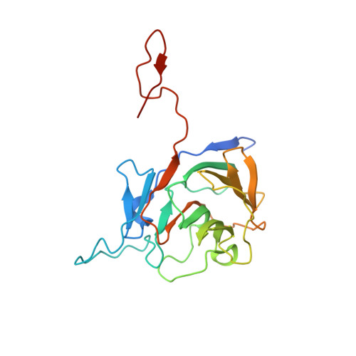
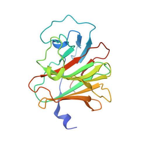
Phosphoantigens glue butyrophilin 3A1 and 2A1 to activate V gamma 9V delta 2 T cells.

Yuan, L.Ma, X.Yang, Y.Qu, Y.Li, X.Zhu, X.Ma, W.Duan, J.Xue, J.Yang, H.Huang, J.W.Yi, S.Zhang, M.Cai, N.Zhang, L.Ding, Q.Lai, K.Liu, C.Zhang, L.Liu, X.Yao, Y.Zhou, S.Li, X.Shen, P.Chang, Q.Malwal, S.R.He, Y.Li, W.Chen, C.Chen, C.C.Oldfield, E.Guo, R.T.Zhang, Y.

(2023) Nature 621: 840-848

  • DOI: https://doi.org/10.1038/s41586-023-06525-3
  • Primary Citation of Related Structures:  
    8HJT, 8IGT, 8IH4, 8IXV, 8IZE, 8IZG, 8JY9, 8JYA, 8JYB, 8JYC, 8JYE, 8JYF

  • PubMed Abstract: 

    In both cancer and infections, diseased cells are presented to human Vγ9Vδ2 T cells through an 'inside out' signalling process whereby structurally diverse phosphoantigen (pAg) molecules are sensed by the intracellular domain of butyrophilin BTN3A1 1-4 . Here we show how-in both humans and alpaca-multiple pAgs function as 'molecular glues' to promote heteromeric association between the intracellular domains of BTN3A1 and the structurally similar butyrophilin BTN2A1. X-ray crystallography studies visualized that engagement of BTN3A1 with pAgs forms a composite interface for direct binding to BTN2A1, with various pAg molecules each positioned at the centre of the interface and gluing the butyrophilins with distinct affinities. Our structural insights guided mutagenesis experiments that led to disruption of the intracellular BTN3A1-BTN2A1 association, abolishing pAg-mediated Vγ9Vδ2 T cell activation. Analyses using structure-based molecular-dynamics simulations, 19 F-NMR investigations, chimeric receptor engineering and direct measurement of intercellular binding force revealed how pAg-mediated BTN2A1 association drives BTN3A1 intracellular fluctuations outwards in a thermodynamically favourable manner, thereby enabling BTN3A1 to push off from the BTN2A1 ectodomain to initiate T cell receptor-mediated γδ T cell activation. Practically, we harnessed the molecular-glue model for immunotherapeutics design, demonstrating chemical principles for developing both small-molecule activators and inhibitors of human γδ T cell function.


  • Organizational Affiliation

    Tsinghua-Peking Center for Life Sciences, State Key Laboratory of Membrane Biology, School of Pharmaceutical Sciences, Tsinghua University, Beijing, China.


Macromolecules
Find similar proteins by:  (by identity cutoff)  |  3D Structure
Entity ID: 1
MoleculeChains Sequence LengthOrganismDetailsImage
Butyrophilin subfamily 2 member A1
A, B
218Homo sapiensMutation(s): 0 
Gene Names: BTN2A1BT2.1BTF1
UniProt & NIH Common Fund Data Resources
Find proteins for Q7KYR7 (Homo sapiens)
Explore Q7KYR7 
Go to UniProtKB:  Q7KYR7
PHAROS:  Q7KYR7
GTEx:  ENSG00000112763 
Entity Groups  
Sequence Clusters30% Identity50% Identity70% Identity90% Identity95% Identity100% Identity
UniProt GroupQ7KYR7
Sequence Annotations
Expand
  • Reference Sequence
Find similar proteins by:  (by identity cutoff)  |  3D Structure
Entity ID: 2
MoleculeChains Sequence LengthOrganismDetailsImage
Butyrophilin subfamily 3 member A1
C, D
196Homo sapiensMutation(s): 0 
Gene Names: BTN3A1
UniProt & NIH Common Fund Data Resources
Find proteins for O00481 (Homo sapiens)
Explore O00481 
Go to UniProtKB:  O00481
PHAROS:  O00481
GTEx:  ENSG00000026950 
Entity Groups  
Sequence Clusters30% Identity50% Identity70% Identity90% Identity95% Identity100% Identity
UniProt GroupO00481
Sequence Annotations
Expand
  • Reference Sequence
Small Molecules
Ligands 6 Unique
IDChains Name / Formula / InChI Key2D Diagram3D Interactions
DMA (Subject of Investigation/LOI)
Query on DMA

Download Ideal Coordinates CCD File 
T [auth C],
Y [auth D]
DIMETHYLALLYL DIPHOSPHATE
C5 H12 O7 P2
CBIDRCWHNCKSTO-UHFFFAOYSA-N
1PE
Query on 1PE

Download Ideal Coordinates CCD File 
F [auth A],
G [auth A],
H [auth A],
V [auth C]
PENTAETHYLENE GLYCOL
C10 H22 O6
JLFNLZLINWHATN-UHFFFAOYSA-N
PG4
Query on PG4

Download Ideal Coordinates CCD File 
I [auth A],
J [auth A],
W [auth C]
TETRAETHYLENE GLYCOL
C8 H18 O5
UWHCKJMYHZGTIT-UHFFFAOYSA-N
PGE
Query on PGE

Download Ideal Coordinates CCD File 
K [auth A]TRIETHYLENE GLYCOL
C6 H14 O4
ZIBGPFATKBEMQZ-UHFFFAOYSA-N
PEG
Query on PEG

Download Ideal Coordinates CCD File 
M [auth B],
N [auth B],
S [auth B]
DI(HYDROXYETHYL)ETHER
C4 H10 O3
MTHSVFCYNBDYFN-UHFFFAOYSA-N
EDO
Query on EDO

Download Ideal Coordinates CCD File 
E [auth A]
L [auth A]
O [auth B]
P [auth B]
Q [auth B]
E [auth A],
L [auth A],
O [auth B],
P [auth B],
Q [auth B],
R [auth B],
U [auth C],
X [auth C]
1,2-ETHANEDIOL
C2 H6 O2
LYCAIKOWRPUZTN-UHFFFAOYSA-N
Experimental Data & Validation

Experimental Data

  • Method: X-RAY DIFFRACTION
  • Resolution: 2.29 Å
  • R-Value Free: 0.216 
  • R-Value Work: 0.171 
  • R-Value Observed: 0.173 
  • Space Group: P 41
Unit Cell:
Length ( Å )Angle ( ˚ )
a = 90.153α = 90
b = 90.153β = 90
c = 167.918γ = 90
Software Package:
Software NamePurpose
REFMACrefinement
HKL-2000data scaling
PDB_EXTRACTdata extraction
HKL-2000data reduction
PHASERphasing

Structure Validation

View Full Validation Report


Ligand Structure Quality Assessment 


Entry History & Funding Information

Deposition Data


Funding OrganizationLocationGrant Number
National Natural Science Foundation of China (NSFC)China32100711

Revision History  (Full details and data files)

  • Version 1.0: 2023-09-13
    Type: Initial release
  • Version 1.1: 2023-09-20
    Changes: Database references
  • Version 1.2: 2023-10-11
    Changes: Database references
  • Version 1.3: 2023-10-18
    Changes: Database references