


Crystal structure of Chitoporin from Vibrio harveyi in complex with gentamicin c1a (multiple binding sites)
Sanram, S., Suginta, W.To be published.
Experimental Data Snapshot
Starting Model: experimental
View more details
Entity ID: 1 | |||||
|---|---|---|---|---|---|
| Molecule | Chains | Sequence Length | Organism | Details | Image |
| Chitoporin | 350 | Vibrio harveyi | Mutation(s): 0 Gene Names: chiP, AL538_13355, VCHENC02_0932 Membrane Entity: Yes | 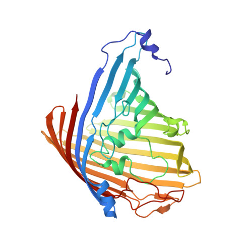 | |
UniProt | |||||
Entity Groups | |||||
| Sequence Clusters | 30% Identity50% Identity70% Identity90% Identity95% Identity100% Identity | ||||
| UniProt Group | L0RVU0 | ||||
Sequence AnnotationsExpand | |||||
Reference Sequence | |||||
| Ligands 3 Unique | |||||
|---|---|---|---|---|---|
| ID | Chains | Name / Formula / InChI Key | 2D Diagram | 3D Interactions | |
| LLL Download:Ideal Coordinates CCD File | H [auth A] | (2R,3R,4R,5R)-2-((1S,2S,3R,4S,6R)-4,6-DIAMINO-3-((2R,3R,6S)-3-AMINO-6-(AMINOMETHYL)-TETRAHYDRO-2H-PYRAN-2-YLOXY)-2-HYDR
OXYCYCLOHEXYLOXY)-5-METHYL-4-(METHYLAMINO)-TETRAHYDRO-2H-PYRAN-3,5-DIOL C19 H39 N5 O7 VEGXETMJINRLTH-BOZYPMBZSA-N |  | ||
| C8E (Subject of Investigation/LOI) Download:Ideal Coordinates CCD File | D [auth A], I [auth B], M [auth C] | (HYDROXYETHYLOXY)TRI(ETHYLOXY)OCTANE C16 H34 O5 FEOZZFHAVXYAMB-UHFFFAOYSA-N |  | ||
| NA Download:Ideal Coordinates CCD File | E [auth A] F [auth A] G [auth A] J [auth B] K [auth B] | SODIUM ION Na FKNQFGJONOIPTF-UHFFFAOYSA-N |  | ||
| Length ( Å ) | Angle ( ˚ ) |
|---|---|
| a = 86.326 | α = 90 |
| b = 134.103 | β = 90 |
| c = 145.787 | γ = 90 |
| Software Name | Purpose |
|---|---|
| PHENIX | refinement |
| HKL-2000 | data scaling |
| HKL-2000 | data reduction |
| PHENIX | phasing |
| Funding Organization | Location | Grant Number |
|---|---|---|
| Vidyasirimedhi Institute of Science and Technology (VISTEC) | Thailand | -- |


