8JSR | pdb_00008jsr

Cryo-EM structure of the anamorelin-bound ghrelin receptor and Gq complex


Experimental Data Snapshot

  • Method: ELECTRON MICROSCOPY
  • Resolution: 2.90 Å
  • Aggregation State: PARTICLE 
  • Reconstruction Method: SINGLE PARTICLE 

wwPDB Validation 3D Report Full Report

Validation slider image for 8JSR

This is version 1.4 of the entry. See complete history

Literature

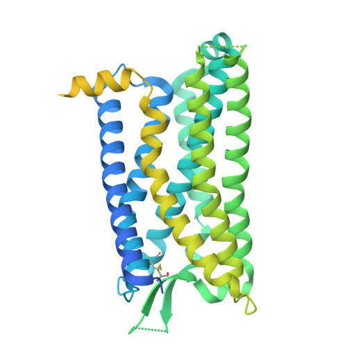
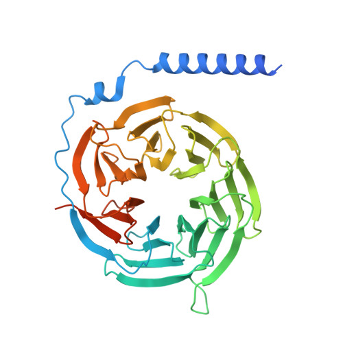
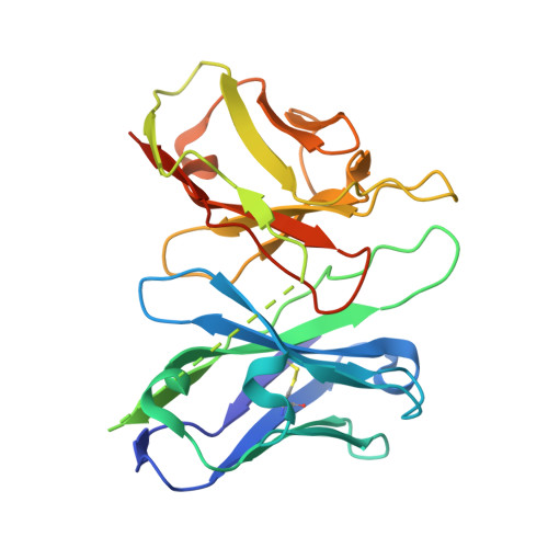
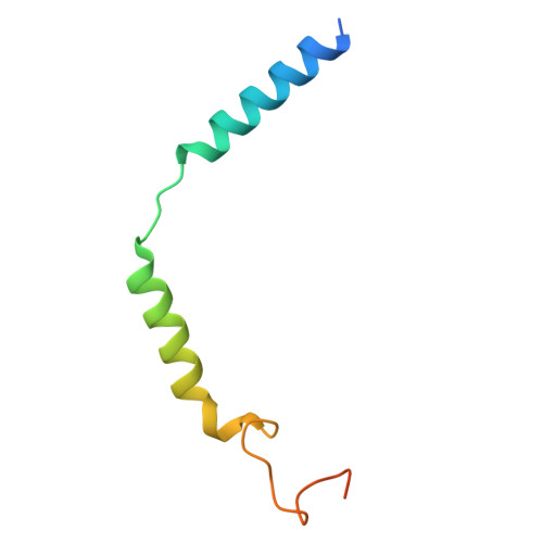
The structure and function of the ghrelin receptor coding for drug actions.

Shiimura, Y.Im, D.Tany, R.Asada, H.Kise, R.Kurumiya, E.Wakasugi-Masuho, H.Yasuda, S.Matsui, K.Kishikawa, J.I.Kato, T.Murata, T.Kojima, M.Iwata, S.Masuho, I.

(2025) Nat Struct Mol Biol 32: 531-542

  • DOI: https://doi.org/10.1038/s41594-024-01481-6
  • Primary Citation Related Structures: 
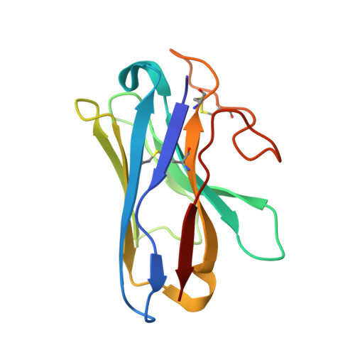
    8JSR

  • PubMed Abstract: 

    Drugs targeting the ghrelin receptor hold therapeutic potential in anorexia, obesity and diabetes. However, developing effective drugs is challenging. To tackle this common issue across a broad drug target, this study aims to understand how anamorelin, the only approved drug targeting the ghrelin receptor, operates compared to other synthetic drugs. Our research elucidated the receptor's structure with anamorelin and miniG q , unveiling anamorelin's superagonistic activity. We demonstrated that ligands with distinct chemical structures uniquely bind to the receptor, resulting in diverse conformations and biasing signal transduction. Moreover, our study showcased the utility of structural information in effectively identifying natural genetic variations altering drug action and causing severe functional deficiencies, offering a basis for selecting the right medication on the basis of the individual's genomic sequence. Thus, by building on structural analysis, this study enhances the foundational framework for selecting therapeutic agents targeting the ghrelin receptor, by effectively leveraging signaling bias and genetic variations.


  • Organizational Affiliation
    • Division of Molecular Genetics, Institute of Life Science, Kurume University, Fukuoka, Japan. shiimura_yuuki@kurume-u.ac.jp.

Macromolecule Content 

  • Total Structure Weight: 193.04 kDa 
  • Atom Count: 10,001 
  • Modeled Residue Count: 1,275 
  • Deposited Residue Count: 1,731 
  • Unique protein chains: 6

Macromolecules

Find similar proteins by:|  3D Structure
Entity ID: 1
MoleculeChains  Sequence LengthOrganismDetailsImage
Growth hormone secretagogue receptor type 1A [auth R]523Homo sapiensMutation(s): 0 
Gene Names: GHSR
Membrane Entity: Yes 
UniProt & NIH Common Fund Data Resources
Find proteins for Q92847 (Homo sapiens)
Explore Q92847 
Go to UniProtKB:  Q92847
PHAROS:  Q92847
GTEx:  ENSG00000121853 
Entity Groups
Sequence Clusters30% Identity50% Identity70% Identity90% Identity95% Identity100% Identity
UniProt GroupQ92847
Sequence Annotations
Expand
Reference Sequence
Find similar proteins by:|  3D Structure
Entity ID: 2
MoleculeChains  Sequence LengthOrganismDetailsImage
Engineered G-alpha-qB [auth A]361Homo sapiensMutation(s): 0 
Find similar proteins by:|  3D Structure
Entity ID: 3
MoleculeChains  Sequence LengthOrganismDetailsImage
Guanine nucleotide-binding protein G(I)/G(S)/G(T) subunit beta-1C [auth B]388Homo sapiensMutation(s): 0 
Gene Names: GNB1
UniProt & NIH Common Fund Data Resources
Find proteins for P62873 (Homo sapiens)
Explore P62873 
Go to UniProtKB:  P62873
PHAROS:  P62873
GTEx:  ENSG00000078369 
Entity Groups
Sequence Clusters30% Identity50% Identity70% Identity90% Identity95% Identity100% Identity
UniProt GroupP62873
Sequence Annotations
Expand
Reference Sequence
Find similar proteins by:|  3D Structure
Entity ID: 4
MoleculeChains  Sequence LengthOrganismDetailsImage
scFv16D [auth C]259Mus musculusMutation(s): 0 
Find similar proteins by:|  3D Structure
Entity ID: 5
MoleculeChains  Sequence LengthOrganismDetailsImage
Guanine nucleotide-binding protein G(I)/G(S)/G(O) subunit gamma-2E [auth G]71Homo sapiensMutation(s): 0 
Gene Names: GNG2
Membrane Entity: Yes 
UniProt & NIH Common Fund Data Resources
Find proteins for P59768 (Homo sapiens)
Explore P59768 
Go to UniProtKB:  P59768
PHAROS:  P59768
GTEx:  ENSG00000186469 
Entity Groups
Sequence Clusters30% Identity50% Identity70% Identity90% Identity95% Identity100% Identity
UniProt GroupP59768
Sequence Annotations
Expand
Reference Sequence
Find similar proteins by:|  3D Structure
Entity ID: 6
MoleculeChains  Sequence LengthOrganismDetailsImage
Nb35F [auth N]129Lama glamaMutation(s): 0 

Small Molecules

Ligands 1 Unique
IDChains Name / Formula / InChI Key2D Diagram3D Interactions
UYI

Query on UYI


Download:Ideal Coordinates CCD File
G [auth R]Anamorelin
C31 H42 N6 O3
VQPFSIRUEPQQPP-MXBOTTGLSA-N

Experimental Data & Validation

Experimental Data

  • Method: ELECTRON MICROSCOPY
  • Resolution: 2.90 Å
  • Aggregation State: PARTICLE 
  • Reconstruction Method: SINGLE PARTICLE 
EM Software:
TaskSoftware PackageVersion
MODEL REFINEMENTPHENIX1.21rc1_5109:

Structure Validation

View Full Validation Report


Entry History 

& Funding Information

Deposition Data


Funding OrganizationLocationGrant Number
Japan Society for the Promotion of Science (JSPS)Japan18K16217, 20K06531, 20KK0359, 22KK0099

Revision History  (Full details and data files)

  • Version 1.0: 2025-01-22
    Type: Initial release
  • Version 1.1: 2025-02-05
    Changes: Data collection, Database references
  • Version 1.2: 2025-04-02
    Changes: Data collection, Database references
  • Version 1.3: 2025-11-26
    Changes: Data collection, Derived calculations, Structure summary
  • Version 1.4: 2025-12-10
    Changes: Data collection, Structure summary