8JPF | pdb_00008jpf

Focused refiment structure of NTSR1 in NTSR1-GRK2-Galpha(q) complexes


Experimental Data Snapshot

  • Method: ELECTRON MICROSCOPY
  • Resolution: 3.02 Å
  • Aggregation State: PARTICLE 
  • Reconstruction Method: SINGLE PARTICLE 

wwPDB Validation 3D Report Full Report

Validation slider image for 8JPF

This is version 1.2 of the entry. See complete history. 

Literature

GPCR activation and GRK2 assembly by a biased intracellular agonist.

Duan, J., Liu, H., Zhao, F., Yuan, Q., Ji, Y., Cai, X., He, X., Li, X., Li, J., Wu, K., Gao, T., Zhu, S., Lin, S., Wang, M.W., Cheng, X., Yin, W., Jiang, Y., Yang, D., Xu, H.E.

(2023) Nature 620: 676-681

  • DOI: https://doi.org/10.1038/s41586-023-06395-9
  • Primary Citation Related Structures: 
    8JPB, 8JPC, 8JPD, 8JPE, 8JPF

  • PubMed Abstract: 

    Phosphorylation of G-protein-coupled receptors (GPCRs) by GPCR kinases (GRKs) desensitizes G-protein signalling and promotes arrestin signalling, which is also modulated by biased ligands 1-6 . The molecular assembly of GRKs on GPCRs and the basis of GRK-mediated biased signalling remain largely unknown owing to the weak GPCR-GRK interactions. Here we report the complex structure of neurotensin receptor 1 (NTSR1) bound to GRK2, Gα q and the arrestin-biased ligand SBI-553 7 . The density map reveals the arrangement of the intact GRK2 with the receptor, with the N-terminal helix of GRK2 docking into the open cytoplasmic pocket formed by the outward movement of the receptor transmembrane helix 6, analogous to the binding of the G protein to the receptor. SBI-553 binds at the interface between GRK2 and NTSR1 to enhance GRK2 binding. The binding mode of SBI-553 is compatible with arrestin binding but clashes with the binding of Gα q protein, thus providing a mechanism for its arrestin-biased signalling capability. In sum, our structure provides a rational model for understanding the details of GPCR-GRK interactions and GRK2-mediated biased signalling.


  • Organizational Affiliation: 
    • State Key Laboratory of Drug Research, Shanghai Institute of Materia Medica, Chinese Academy of Sciences, Shanghai, China. duanjia@simm.ac.cn.

Macromolecule Content 

  • Total Structure Weight: 47.58 kDa 
  • Atom Count: 2,269 
  • Modeled Residue Count: 304 
  • Deposited Residue Count: 424 
  • Unique protein chains: 2

Macromolecules

Find similar proteins by:  Sequence   |   3D Structure  
Entity ID: 1
MoleculeChains  Sequence LengthOrganismDetailsImage
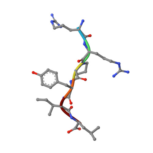
NTSA [auth L]6synthetic constructMutation(s): 0 
Sequence Annotations
Expand
Reference Sequence
Find similar proteins by:|  3D Structure
Entity ID: 2
MoleculeChains  Sequence LengthOrganismDetailsImage
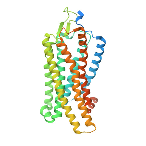
Neurotensin receptor type 1B [auth R]418Homo sapiensMutation(s): 0 
Gene Names: NTSR1, NTRR
Membrane Entity: Yes 
UniProt & NIH Common Fund Data Resources
Find proteins for P30989 (Homo sapiens)
Explore P30989 
Go to UniProtKB:  P30989
PHAROS:  P30989
GTEx:  ENSG00000101188 
Entity Groups
Sequence Clusters30% Identity50% Identity70% Identity90% Identity95% Identity100% Identity
UniProt GroupP30989
Sequence Annotations
Expand
Reference Sequence

Small Molecules

Ligands 1 Unique
IDChains Name / Formula / InChI Key2D Diagram3D Interactions
SRW

Query on SRW


Download:Ideal Coordinates CCD File
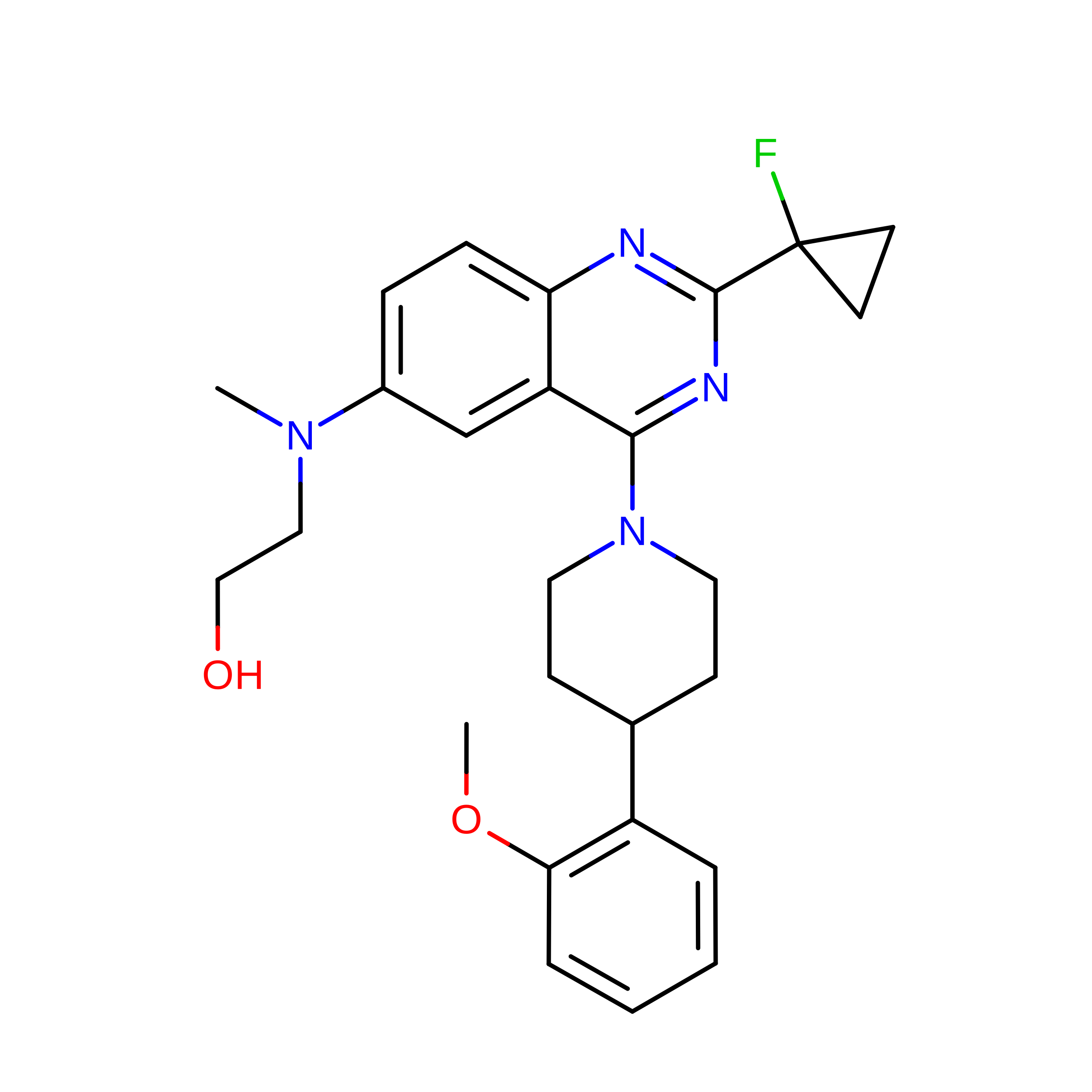
C [auth R]2-[{2-(1-fluorocyclopropyl)-4-[4-(2-methoxyphenyl)piperidin-1-yl]quinazolin-6-yl}(methyl)amino]ethan-1-ol
C26 H31 F N4 O2
BLWXTJQMEBQCIZ-UHFFFAOYSA-N
Binding Affinity Annotations 
IDSourceBinding Affinity
SRW BindingDB:  8JPF EC50: min: 1.2, max: 500 (nM) from 4 assay(s)

Experimental Data & Validation

Experimental Data

  • Method: ELECTRON MICROSCOPY
  • Resolution: 3.02 Å
  • Aggregation State: PARTICLE 
  • Reconstruction Method: SINGLE PARTICLE 

Structure Validation

View Full Validation Report


Entry History 

& Funding Information

Deposition Data


Funding OrganizationLocationGrant Number
National Natural Science Foundation of China (NSFC)China--

Revision History  (Full details and data files)

  • Version 1.0: 2023-08-09
    Type: Initial release
  • Version 1.1: 2023-08-16
    Changes: Data collection, Database references
  • Version 1.2: 2023-08-30
    Changes: Database references