Structural and functional insights into the lipid regulation of human anion exchanger 2.
Zhang, W., Ding, D., Lu, Y., Chen, H., Jiang, P., Zuo, P., Wang, G., Luo, J., Yin, Y., Luo, J., Yin, Y.(2024) Nat Commun 15: 759-759
- PubMed: 38272905 Search on PubMedSearch on PubMed Central
- DOI: https://doi.org/10.1038/s41467-024-44966-0
- Primary Citation Related Structures:
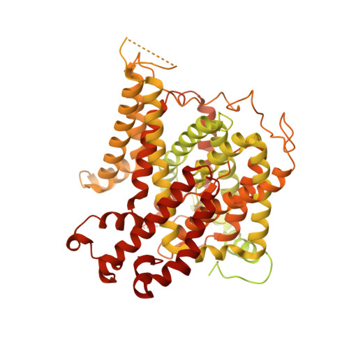
8JNI, 8JNJ - PubMed Abstract:
Anion exchanger 2 (AE2) is an electroneutral Na + -independent Cl - /HCO 3 - exchanger belongs to the SLC4 transporter family. The widely expressed AE2 participates in a variety of physiological processes, including transepithelial acid-base secretion and osteoclastogenesis. Both the transmembrane domains (TMDs) and the N-terminal cytoplasmic domain (NTD) are involved in regulation of AE2 activity. However, the regulatory mechanism remains unclear. Here, we report a 3.2 Å cryo-EM structure of the AE2 TMDs in complex with PIP 2 and a 3.3 Å full-length mutant AE2 structure in the resting state without PIP 2 . We demonstrate that PIP 2 at the TMD dimer interface is involved in the substrate exchange process. Mutation in the PIP 2 binding site leads to the displacement of TM7 and further stabilizes the interaction between the TMD and the NTD. Reduced substrate transport activity and conformation similar to AE2 in acidic pH indicating the central contribution of PIP 2 to the function of AE2.
- Institute of Systems Biomedicine, Department of Pathology, Beijing Key Laboratory of Tumor Systems Biology, Peking-Tsinghua Center for Life Sciences, School of Basic Medical Sciences, Peking University Health Science Center, Beijing, 100191, China.
Organizational Affiliation: