8JMX | pdb_00008jmx

The crystal structure of human aurka kinase domain in complex with AURKA-A2


Experimental Data Snapshot

  • Method: X-RAY DIFFRACTION
  • Resolution: 2.95 Å
  • R-Value Free: 
    0.306 (Depositor), 0.300 (DCC) 
  • R-Value Work: 
    0.222 (Depositor), 0.230 (DCC) 
  • R-Value Observed: 
    0.231 (Depositor) 

Starting Model: experimental
View more details

wwPDB Validation 3D Report Full Report

Validation slider image for 8JMX

Ligand Structure Quality Assessment 


This is version 1.1 of the entry. See complete history

Literature

Multitarget inhibitors/probes that target LRRK2 and AURORA A kinases noncovalently and covalently.

Wang, W.Wang, X.Tang, G.Zhu, C.Xiang, M.Xiao, Q.Zhang, Z.M.Gao, L.Yao, S.Q.

(2023) Chem Commun (Camb) 59: 10789-10792

  • DOI: https://doi.org/10.1039/d3cc03530a
  • Primary Citation Related Structures: 
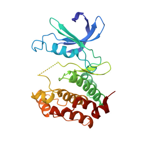
    8JMX

  • PubMed Abstract: 

    Herein, we report a salicylaldehyde-based, reversible covalent inhibitor (A2) that possesses moderate cellular activity against AURKA with a prolonged residence time and shows significant non-covalent inhibition towards LRRK2. Our results indicated that this multitarget kinase inhibitor may be used as the starting point for future development of more potent, selective and dual-targeting covalent kinase inhibitors against AURKA and LRRK2 for mitophagy.


  • Organizational Affiliation
    • School of Pharmaceutical Sciences (Shenzhen), Shenzhen Campus of Sun Yat-sen University, Shenzhen 518000, China. gaolq@mail.sysu.edu.cn.

Macromolecule Content 

  • Total Structure Weight: 30.89 kDa 
  • Atom Count: 1,956 
  • Modeled Residue Count: 250 
  • Deposited Residue Count: 263 
  • Unique protein chains: 1

Macromolecules

Find similar proteins by:|  3D Structure
Entity ID: 1
MoleculeChains  Sequence LengthOrganismDetailsImage
Aurora kinase A263Homo sapiensMutation(s): 0 
Gene Names: AURKAAIKAIRK1ARK1AURAAYK1BTAKIAK1STK15STK6
EC: 2.7.11.1
UniProt & NIH Common Fund Data Resources
Find proteins for O14965 (Homo sapiens)
Explore O14965 
Go to UniProtKB:  O14965
PHAROS:  O14965
GTEx:  ENSG00000087586 
Entity Groups
Sequence Clusters30% Identity50% Identity70% Identity90% Identity95% Identity100% Identity
UniProt GroupO14965
Sequence Annotations
Expand
Reference Sequence

Small Molecules

Ligands 1 Unique
IDChains Name / Formula / InChI Key2D Diagram3D Interactions
E47
(Subject of Investigation/LOI)

Query on E47


Download:Ideal Coordinates CCD File
B [auth A]5-(4-morpholin-4-yl-7H-pyrrolo[2,3-d]pyrimidin-5-yl)-2-oxidanyl-benzaldehyde
C17 H16 N4 O3
OPQSSAIBYNXUAR-UHFFFAOYSA-N

Experimental Data & Validation

Experimental Data

  • Method: X-RAY DIFFRACTION
  • Resolution: 2.95 Å
  • R-Value Free:  0.306 (Depositor), 0.300 (DCC) 
  • R-Value Work:  0.222 (Depositor), 0.230 (DCC) 
  • R-Value Observed: 0.231 (Depositor) 
Space Group: P 61 2 2
Unit Cell:
Length ( Å )Angle ( ˚ )
a = 83.133α = 90
b = 83.133β = 90
c = 171.452γ = 120
Software Package:
Software NamePurpose
PHENIXrefinement
autoPROCdata reduction
XDSdata scaling
PHENIXphasing

Structure Validation

View Full Validation Report


Ligand Structure Quality Assessment 


Entry History 

& Funding Information

Deposition Data


Funding OrganizationLocationGrant Number
Not funded--

Revision History  (Full details and data files)

  • Version 1.0: 2023-10-18
    Type: Initial release
  • Version 1.1: 2024-11-13
    Changes: Structure summary