8JHW | pdb_00008jhw

The first crystal structure of a H-2Kb-restricted decapeptide from Cryptosporidium parvum


Experimental Data Snapshot

  • Method: X-RAY DIFFRACTION
  • Resolution: 3.12 Å
  • R-Value Free: 
    0.288 (Depositor), 0.288 (DCC) 
  • R-Value Work: 
    0.245 (Depositor), 0.245 (DCC) 

Starting Model: experimental
View more details

wwPDB Validation 3D Report Full Report

Validation slider image for 8JHW

This is version 1.2 of the entry. See complete history

Literature

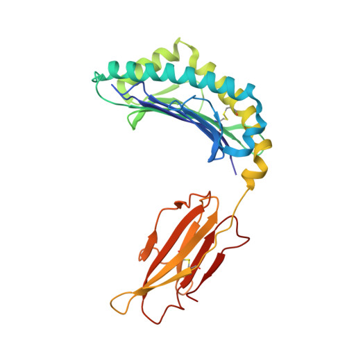
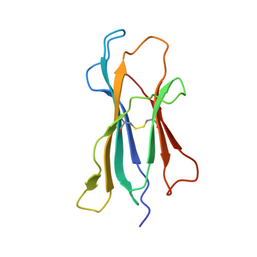
Structural analyses of Cryptosporidium parvum epitopes reveal a novel scheme of decapeptide binding to H-2K b.

Wang, Y.Chang, Y.Yin, F.Kang, C.Meng, Y.Xu, F.Liu, Y.Zhang, Y.Wu, C.Fan, S.Zhao, J.

(2025) J Struct Biol 217: 108168-108168

  • DOI: https://doi.org/10.1016/j.jsb.2025.108168
  • Primary Citation Related Structures: 
    8JHW

  • PubMed Abstract: 

    Cryptosporidium has gained much attention as a major cause of diarrhea worldwide. Here, we present the first structure of H-2K b complexed with a decapeptide from Cryptosporidium parvum Gp40/15 protein (Gp40/15-VTF10). In contrast to all published structures, the aromatic residue P3-Phe of Gp40/15-VTF10 is anchored in pocket C rather than the canonical Y/F at P5 or P6 reported for octapeptides and nonapeptides. The results of in vitro refolding assays and circular dichroism experiments showed that the side chains of P3 and P5 play key roles in Gp40/15-VTF10 peptide binding. However, functional analysis of decapeptide epitopes revealed that the Gp40/15-VTF10 peptide did not elicit a strong CD8 + T immune response, whereas the decapeptide epitope MEDLE2-INF10 induced a significant CD8 + T-cell response in peptide-immunized C57BL/6 mice. Using a model structure of H-2K b -INF10 complex, we found that the antigenic decapeptide INF10 exhibits a completely different conformation, with the aromatic anchors P3F and P7F docked into the D and C pockets, respectively, while similar peptide conformation and hydrogen bond interactions between the peptide and major histocompatibility complex were found in the resolved H-2K b -SVF9 complex. As the H-2K b molecule predominantly prefers octapeptides with a strong anchor of P5 Y/F (or P6 Y/F for nonapeptides) binding to the C pocket, we propose that P7 Y/F in the C pocket may represent a novel binding mode for decapeptides. The results should increase the accuracy of T-cell epitope prediction and support the development of T-cell epitope vaccines against cryptosporidiosis.


  • Organizational Affiliation
    • State Key Laboratory of Agricultural Microbiology, College of Veterinary Medicine, Huazhong Agricultural University, Wuhan, PR China; College of Life Science and Agronomy, Zhoukou Normal University, Zhoukou, PR China.

Macromolecule Content 

  • Total Structure Weight: 45.01 kDa 
  • Atom Count: 3,159 
  • Modeled Residue Count: 384 
  • Deposited Residue Count: 385 
  • Unique protein chains: 3

Macromolecules

Find similar proteins by:|  3D Structure
Entity ID: 1
MoleculeChains  Sequence LengthOrganismDetailsImage
H-2 class I histocompatibility antigen, K-B alpha chain275Mus musculusMutation(s): 0 
Gene Names: H2-K1H2-K
UniProt
Find proteins for P01901 (Mus musculus)
Explore P01901 
Go to UniProtKB:  P01901
Entity Groups
Sequence Clusters30% Identity50% Identity70% Identity90% Identity95% Identity100% Identity
UniProt GroupP01901
Sequence Annotations
Expand
Reference Sequence
Find similar proteins by:|  3D Structure
Entity ID: 2
MoleculeChains  Sequence LengthOrganismDetailsImage
Beta-2-microglobulin100Mus musculusMutation(s): 0 
Gene Names: B2m
UniProt & NIH Common Fund Data Resources
Find proteins for P01887 (Mus musculus)
Explore P01887 
Go to UniProtKB:  P01887
IMPC:  MGI:88127
Entity Groups
Sequence Clusters30% Identity50% Identity70% Identity90% Identity95% Identity100% Identity
UniProt GroupP01887
Sequence Annotations
Expand
Reference Sequence
Find similar proteins by:  Sequence   |   3D Structure  
Entity ID: 3
MoleculeChains  Sequence LengthOrganismDetailsImage
VAL-THR-PHE-GLU-LYS-SER-TYR-ASN-THR-VAL10Cryptosporidium parvumMutation(s): 0 
UniProt
Find proteins for A0A7S7LF56 (Cryptosporidium parvum)
Explore A0A7S7LF56 
Go to UniProtKB:  A0A7S7LF56
Entity Groups
Sequence Clusters30% Identity50% Identity70% Identity90% Identity95% Identity100% Identity
UniProt GroupA0A7S7LF56
Sequence Annotations
Expand
Reference Sequence

Experimental Data & Validation

Experimental Data

  • Method: X-RAY DIFFRACTION
  • Resolution: 3.12 Å
  • R-Value Free:  0.288 (Depositor), 0.288 (DCC) 
  • R-Value Work:  0.245 (Depositor), 0.245 (DCC) 
Space Group: P 65 2 2
Unit Cell:
Length ( Å )Angle ( ˚ )
a = 86.94α = 90
b = 86.94β = 90
c = 293.73γ = 120
Software Package:
Software NamePurpose
REFMACrefinement
HKL-3000data reduction
HKL-3000data scaling
PHASERphasing

Structure Validation

View Full Validation Report


Entry History 

& Funding Information

Deposition Data

  • Released Date: 2024-05-29 
  • Deposition Author(s): Fan, S., Wang, Y.
  • This entry supersedes: 8JHV

Funding OrganizationLocationGrant Number
National Science Foundation (NSF, China)China31702232

Revision History  (Full details and data files)

  • Version 1.0: 2024-05-29
    Type: Initial release
  • Version 1.1: 2024-11-13
    Changes: Structure summary
  • Version 1.2: 2025-12-10
    Changes: Advisory, Database references