8JEO | pdb_00008jeo

Crystal structure of TIGIT in complexed with Tiragolumab


Experimental Data Snapshot

  • Method: X-RAY DIFFRACTION
  • Resolution: 2.06 Å
  • R-Value Free: 
    0.205 (Depositor), 0.211 (DCC) 
  • R-Value Work: 
    0.172 (Depositor), 0.178 (DCC) 
  • R-Value Observed: 
    0.174 (Depositor) 

Starting Models: experimental
View more details

wwPDB Validation 3D Report Full Report

Validation slider image for 8JEO

This is version 1.3 of the entry. See complete history

Literature

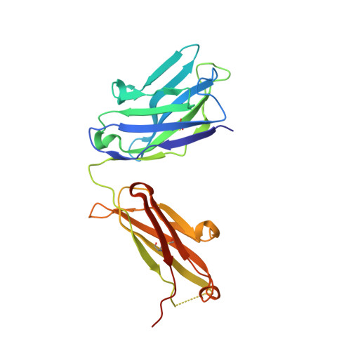
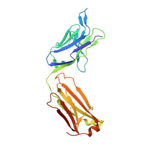
Structural insights into the unique pH-responsive characteristics of the anti-TIGIT therapeutic antibody Ociperlimab.

Sun, J.Zhang, X.Xue, L.Cheng, L.Zhang, J.Chen, X.Shen, Z.Li, K.Wang, L.Huang, C.Song, J.

(2024) Structure 32: 550

  • DOI: https://doi.org/10.1016/j.str.2024.02.009
  • Primary Citation Related Structures: 
    8JEL, 8JEN, 8JEO, 8JEP, 8JEQ

  • PubMed Abstract: 

    TIGIT is mainly expressed on T cells and is an inhibitory checkpoint receptor that binds to its ligand PVR in the tumor microenvironment. Anti-TIGIT monoclonal antibodies (mAbs) such as Ociperlimab and Tiragolumab block the TIGIT-PVR interaction and are in clinical development. However, the molecular blockade mechanism of these mAbs remains elusive. Here, we report the crystal structures of TIGIT in complex with Ociperlimab_Fab and Tiragolumab_Fab revealing that both mAbs bind TIGIT with a large steric clash with PVR. Furthermore, several critical epitopic residues are identified. Interestingly, the binding affinity of Ociperlimab toward TIGIT increases approximately 17-fold when lowering the pH from 7.4 to 6.0. Our structure shows a strong electrostatic interaction between ASP103 HCDR3 and HIS76 TIGIT explaining the pH-responsive mechanism of Ociperlimab. In contrast, Tiragolumab does not show an acidic pH-dependent binding enhancement. Our results provide valuable information that could help to improve the efficacy of therapeutic antibodies for cancer treatment.


  • Organizational Affiliation
    • Department of Biologics, BeiGene (Beijing) Co., Ltd, Beijing, China.

Macromolecule Content 

  • Total Structure Weight: 124.83 kDa 
  • Atom Count: 9,082 
  • Modeled Residue Count: 1,096 
  • Deposited Residue Count: 1,140 
  • Unique protein chains: 3

Macromolecules

Find similar proteins by:|  3D Structure
Entity ID: 1
MoleculeChains  Sequence LengthOrganismDetailsImage
T-cell immunoreceptor with Ig and ITIM domains
A, D
115Homo sapiensMutation(s): 0 
Gene Names: TIGITVSIG9VSTM3
UniProt & NIH Common Fund Data Resources
Find proteins for Q495A1 (Homo sapiens)
Explore Q495A1 
Go to UniProtKB:  Q495A1
PHAROS:  Q495A1
GTEx:  ENSG00000181847 
Entity Groups
Sequence Clusters30% Identity50% Identity70% Identity90% Identity95% Identity100% Identity
UniProt GroupQ495A1
Sequence Annotations
Expand
Reference Sequence
Find similar proteins by:|  3D Structure
Entity ID: 2
MoleculeChains  Sequence LengthOrganismDetailsImage
antibody heavy chain
B, E
235Homo sapiensMutation(s): 0 
Find similar proteins by:|  3D Structure
Entity ID: 3
MoleculeChains  Sequence LengthOrganismDetailsImage
antibody light chain
C, F
220Homo sapiensMutation(s): 0 

Experimental Data & Validation

Experimental Data

  • Method: X-RAY DIFFRACTION
  • Resolution: 2.06 Å
  • R-Value Free:  0.205 (Depositor), 0.211 (DCC) 
  • R-Value Work:  0.172 (Depositor), 0.178 (DCC) 
  • R-Value Observed: 0.174 (Depositor) 
Space Group: I 4
Unit Cell:
Length ( Å )Angle ( ˚ )
a = 214.161α = 90
b = 214.161β = 90
c = 66.885γ = 90
Software Package:
Software NamePurpose
PHENIXrefinement
XDSdata scaling
XDSdata reduction
PHASERphasing

Structure Validation

View Full Validation Report


Entry History 

& Funding Information

Deposition Data


Funding OrganizationLocationGrant Number
Not funded--

Revision History  (Full details and data files)

  • Version 1.0: 2024-02-28
    Type: Initial release
  • Version 1.1: 2024-03-20
    Changes: Database references
  • Version 1.2: 2024-05-22
    Changes: Database references
  • Version 1.3: 2024-11-06
    Changes: Structure summary