


Structure of endo-1,3-fucanase at 1.60 Angstroms resolution.
Chen, G.N., Chang, Y.G., Zhang, Y.Y.To be published.
Experimental Data Snapshot
wwPDB Validation 3D Report Full Report

Entity ID: 1 | |||||
|---|---|---|---|---|---|
| Molecule | Chains | Sequence Length | Organism | Details | Image |
| endo-1,3-fucanase | 529 | Wenyingzhuangia aestuarii | Mutation(s): 0 EC: 3.2.1.211 | 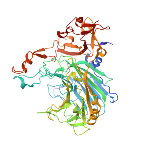 | |
UniProt | |||||
Find proteins for A0AAT8XU97 (Wenyingzhuangia aestuarii) Explore A0AAT8XU97 Go to UniProtKB: A0AAT8XU97 | |||||
Entity Groups | |||||
| Sequence Clusters | 30% Identity50% Identity70% Identity90% Identity95% Identity100% Identity | ||||
| UniProt Group | A0AAT8XU97 | ||||
Sequence AnnotationsExpand | |||||
Reference Sequence | |||||
| Length ( Å ) | Angle ( ˚ ) |
|---|---|
| a = 71.176 | α = 90 |
| b = 70.496 | β = 107.53 |
| c = 103.003 | γ = 90 |
| Software Name | Purpose |
|---|---|
| PHENIX | refinement |
| XDS | data reduction |
| Aimless | data scaling |
| PHASER | phasing |
| Funding Organization | Location | Grant Number |
|---|---|---|
| National Natural Science Foundation of China (NSFC) | China | U22A20542 |


